Review
. 2011 Mar-Apr;17(3-4):281-92.
doi: 10.2119/molmed.2010.00178. Epub 2010 Nov 12.

A paradoxical role for myeloid-derived suppressor cells in sepsis and trauma

Affiliations
Review

A paradoxical role for myeloid-derived suppressor cells in sepsis and trauma

Alex G Cuenca et al. Mol Med. 2011 Mar-Apr.

Abstract

Myeloid-derived suppressor cells (MDSCs) are a heterogenous population of immature myeloid cells whose numbers dramatically increase in chronic and acute inflammatory diseases, including cancer, autoimmune disease, trauma, burns and sepsis. Studied originally in cancer, these cells are potently immunosuppressive, particularly in their ability to suppress antigen-specific CD8(+) and CD4(+) T-cell activation through multiple mechanisms, including depletion of extracellular arginine, nitrosylation of regulatory proteins, and secretion of interleukin 10, prostaglandins and other immunosuppressive mediators. However, additional properties of these cells, including increased reactive oxygen species and inflammatory cytokine production, as well as their universal expansion in nearly all inflammatory conditions, suggest that MDSCs may be more of a normal component of the inflammatory response ("emergency myelopoiesis") than simply a pathological response to a growing tumor. Recent evocative data even suggest that the expansion of MDSCs in acute inflammatory processes, such as burns and sepsis, plays a beneficial role in the host by increasing immune surveillance and innate immune responses. Although clinical efforts are currently underway to suppress MDSC numbers and function in cancer to improve antineoplastic responses, such approaches may not be desirable or beneficial in other clinical conditions in which immune surveillance and antimicrobial activities are required.


Figures
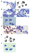
Figure 1
Figure 1
Appearance of MDSCs obtained by cytospin from different clinical conditions. (A) Murine control; (B) murine cancer; (C) human cancer; (D) murine sepsis; (E) murine trauma; (F) murine autoimmunity. GR-1+ splenocytes were obtained from a variety of experimental conditions, including healthy controls (Left, unpublished data; Right [48]), animals with transplantable tumors (Left and Right, unpublished data), human cancer (18), after trauma (48), sepsis (Left [12] and Right [28]) and experimental autoimmune disease (56). Although the photomicrographs were obtained under different experimental conditions, the populations of GR-1+ cells differ dramatically not only within the same condition, but also between conditions. MDSCs phenotypically vary from more mature-appearing neutrophils, to ringed cells, to more immature blast-like–appearing cells. Arrows indicate different subsets of MDSCs: granulocytic (black), ringlike (red), monocytic (orange) and blast-like (green).
Figure 2
Figure 2
Emergency myelopoiesis and the expansion of the MDSC population in sepsis. The figure demonstrates the changes in the early populations of HSCs and multipotent progenitors, which are the progenitors for MDSCs. Although expansion of the MDSC population can be explained in part through increased myelopoiesis, defects in the maturation and differentiation of these populations also contributes to their expansion and their immature phenotype.
Figure 3
Figure 3
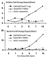
Kinetics of MDSC appearance in the spleen relative to the changes in regulatory T cells and myeloid dendritic cells during sepsis. During sepsis, there are dramatic dynamic changes in the numbers of different regulatory cell populations. In the spleen of mice that survive a polymicrobial sepsis (cecal ligation and puncture), there is a rapid and transient increase in both the relative and absolute numbers of regulatory T cells (CD4+CD25+FoxP3+), with a simultaneous loss of myeloid dendritic cells (CD11b+CD11c+). The response is transient, unlike the increases in both relative and absolute numbers of MDSCs (CD11b+GR-1+) which occur later and are much more sustained.
Figure 4
Figure 4
Expansion of Gr-1+CD11b+ cells in spleen and lymph nodes of burned and infected mice. (A), Flow cytometric scattergram showing populations of GR-1+CD11b+ cells in a spleen from a control mouse. Details concerning the experimental methods are contained in the Supplementary Methods. (B), Flow cytometric scattergram showing expansion of GR-1+CD11b+ cells in a spleen from a mouse that underwent a 10% burn and Pseudomonas infection harvested on d 7. (C), Summary data of the effect of the burn and Pseudomonas infection on splenic GR-1+CD11b+ cells. (D), Summary data of the effect of the burn and Pseudomonas infection on lymph node GR-1+CD11b+ cells. In both lymphoid organs, there was a significant increase in the expansion of GR-1+CD11b+ cells from the Pseudomonas infection alone experimental group as well as the 10% burn + infection group as compared with controls. Values represent the median and estimated quartiles of five animals per group, using a box plot representation, since the values failed tests of normality and equal variance. * indicates that the control group differed only from the groups of mice receiving the Pseudomonas infection, either alone or superimposed on the burn; P < 0.05.
Figure 5
Figure 5
Effect of different approaches to blocking MDSC expansion on survival to sepsis. Mice were pretreated with either gemcitabine or anti–GR-1 monoclonal antibodies, or antibodies to SDF-1 and subjected to an LD30 cecal ligation and puncture, as described in detail in the Supplemental Methods. Survival to CLP was dramatically reduced in mice treated with either gemcitabine (A, n = 20 in each group) or anti–GR-1 antibodies (C, n = 20 in each group). In surviving animals, there was evidence that the expansion of the MDSC population at 7 d was markedly suppressed (*P < 0.01).
Figure 6
Figure 6
MDL1 expression in spleens of patients after trauma or who have died from sepsis. Paraffin-embedded sections from spleens of patients who had either died from sepsis (C and D), or had suffered blunt trauma and undergone splenectomy (B), or had undergone splenectomy for nontrauma or sepsis indications (A) were stained with antibodies to MDL-1. Details about the patient population can be found in (52). Sepsis was associated with the dissolution of lymphoid follicles and the dramatic increase in MDL-1+ cells. Trauma was also associated with more modest increases, although the pattern of MDL-1+ cell expression was markedly different. Staining protocol is provided in the Supplemental Methods. Magnification 100×.

References

    1. Bronte V. Myeloid-derived suppressor cells in inflammation: uncovering cell subsets with enhanced immunosuppressive functions. Eur J Immunol. 2009;39:2670–2. - PubMed
    1. Gabrilovich DI, Nagaraj S. Myeloid-derived suppressor cells as regulators of the immune system. Nat Rev Immunol. 2009;9:162–74. - PMC - PubMed
    1. Ostrand-Rosenberg S, Sinha P. Myeloid-derived suppressor cells: linking inflammation and cancer. J Immunol. 2009;182:4499–506. - PMC - PubMed
    1. Murphey ED, et al. Diminished bacterial clearance is associated with decreased IL-12 and interferon-gamma production but a sustained proinflammatory response in a murine model of postseptic immunosuppression. Shock. 2004;21:415–25. - PubMed
    1. Young MR, Newby M, Wepsic HT. Hematopoiesis and suppressor bone marrow cells in mice bearing large metastatic Lewis lung carcinoma tumors. Cancer Res. 1987;47:100–5. - PubMed

Publication types