Biocompatibility of orthodontic adhesives in rat subcutaneous tissue
- PMID: 21085807
- PMCID: PMC4246383
- DOI: 10.1590/s1678-77572010000500013
Biocompatibility of orthodontic adhesives in rat subcutaneous tissue
Abstract
Objective: The objective of the present study was to verify the hypothesis that no difference in biocompatibility exists between different orthodontic adhesives.
Material and methods: Thirty male Wistar rats were used in this study and divided into five groups (n=6): Group 1 (control, distilled water), Group 2 (Concise), Group 3 (Xeno III), Group 4 (Transbond XT), and Group 5 (Transbond plus Self-Etching Primer). Two cavities were performed in the subcutaneous dorsum of each animal to place a polyvinyl sponge soaked with 2 drops of the respective adhesive in each surgical loci. Two animals of each group were sacrificed after 7, 15, and 30 days, and their tissues were analyzed by using an optical microscope.
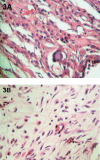
Results: At day 7, Groups 3 (Transbond XT) and 4 (Xeno III) showed intense mono- and polymorphonuclear inflammatory infiltrate with no differences between them, whereas Groups 1 (control) and 2 (Concise) showed moderate mononuclear inflammatory infiltrate. At day 15, severe inflammation was observed in Group 3 (Transbond XT) compared to other groups. At day 30, the same group showed a more expressive mononuclear inflammatory infiltrate compared to other groups.
Conclusion: Among the orthodontic adhesive analyzed, it may be concluded that Transbond XT exhibited the worst biocompatibility. However, one cannot interpret the specificity of the data generated in vivo animal models as a human response.
Figures


Similar articles
-
Morphological and immunohistochemical analysis of the biocompatibility of resin-modified cements.Microsc Res Tech. 2017 May;80(5):504-510. doi: 10.1002/jemt.22822. Epub 2016 Dec 28. Microsc Res Tech. 2017. PMID: 28029189
-
Effect of early orthodontic force on shear bond strength of orthodontic brackets bonded with different adhesive systems.Am J Orthod Dentofacial Orthop. 2010 Aug;138(2):208-14. doi: 10.1016/j.ajodo.2008.09.034. Am J Orthod Dentofacial Orthop. 2010. PMID: 20691363
-
Shear bond strength of metallic orthodontic brackets bonded to enamel prepared with Self-Etching Primer.Angle Orthod. 2005 Sep;75(5):849-53. doi: 10.1043/0003-3219(2005)75[849:SBSOMO]2.0.CO;2. Angle Orthod. 2005. PMID: 16285044
-
Enamel shear bond strength of different primers combined with an orthodontic adhesive paste.Biomed Tech (Berl). 2017 Aug 28;62(4):415-420. doi: 10.1515/bmt-2016-0241. Biomed Tech (Berl). 2017. PMID: 28640749
-
Laboratory evaluation of adhesive systems.Oper Dent. 1992;Suppl 5:50-61. Oper Dent. 1992. PMID: 1470553 Review.
Cited by
-
Bond strength, degree of conversion, and microorganism adhesion using different bracket-to-enamel bonding protocols.J Orofac Orthop. 2023 Oct;84(Suppl 3):210-221. doi: 10.1007/s00056-022-00430-6. Epub 2022 Oct 17. J Orofac Orthop. 2023. PMID: 36251054 English.
-
Evaluation of the genotoxicity and cytotoxicity in the buccal epithelial cells of patients undergoing orthodontic treatment with three light-cured bonding composites by using micronucleus testing.Korean J Orthod. 2014 May;44(3):128-35. doi: 10.4041/kjod.2014.44.3.128. Epub 2014 May 19. Korean J Orthod. 2014. PMID: 24892026 Free PMC article.
-
In Vitro Cytotoxicity Assessment of an Orthodontic Composite Containing Titanium-dioxide Nano-particles.J Dent Res Dent Clin Dent Prospects. 2013 Fall;7(4):192-8. doi: 10.5681/joddd.2013.031. Epub 2013 Dec 18. J Dent Res Dent Clin Dent Prospects. 2013. PMID: 24578816 Free PMC article.
-
Comparison of crevicular fluid cytokine levels after the application of surface sealants : A randomized trial.J Orofac Orthop. 2019 Sep;80(5):242-253. doi: 10.1007/s00056-019-00184-8. Epub 2019 Jul 22. J Orofac Orthop. 2019. PMID: 31444542 Clinical Trial. English.
-
Effect of degree of conversion on in vivo biocompatibility of flowable resin used for bioprotection of mini-implants.Angle Orthod. 2016 Jan;86(1):157-63. doi: 10.2319/112914-856.1. Angle Orthod. 2016. PMID: 26716818 Free PMC article.
References
-
- al-Dawood A, Wennberg A. Biocompatibility of dentin bonding agents. Endod Dent Traumatol. 1993;9(1):1–7. - PubMed
-
- Axford SE, Ogden GR, Stewart AM, Saleh HA, Ross PE, Hopwood D. Fluid phase endocytosis within buccal mucosal cells of alcohol misusers. Oral Oncol. 1999;35(1):86–92. - PubMed
-
- Bouillaguet S, Wataha JC, Hanks CT, Ciucchi B, Holz J. In vitro cytotoxicity and dentin permeability of HEMA. J Endod. 1996;22(5):244–248. - PubMed
-
- Costa CA, Giro EM, Nascimento AB, Teixeira HM, Hebling J. Short-term evaluation of the pulpo-dentin complex response to a resin-modified glass-ionomer cement and a bonding agent applied in deep cavities. Dent Mater. 2003;19(8):739–746. - PubMed
-
- Costa CA, Hebling J, Hanks CT. Current status of pulp capping with dentin adhesive systems: a review. Dent Mater. 2000;16(3):188–197. - PubMed
MeSH terms
Substances
LinkOut - more resources
Full Text Sources

