

Pyogenic granuloma on the upper lip: an unusual location
- PMID: 21085814
- PMCID: PMC4246389
- DOI: 10.1590/s1678-77572010000500019
Pyogenic granuloma on the upper lip: an unusual location
Abstract
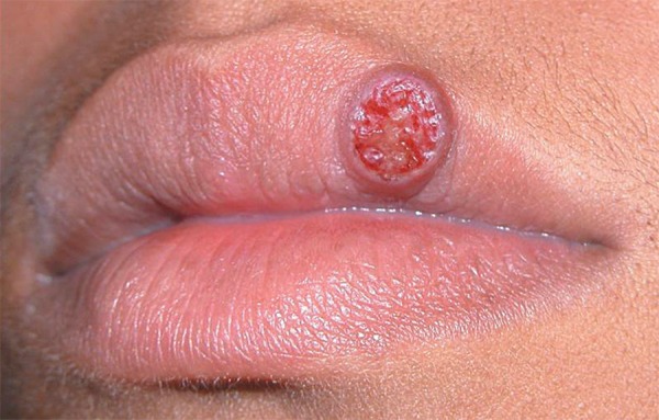
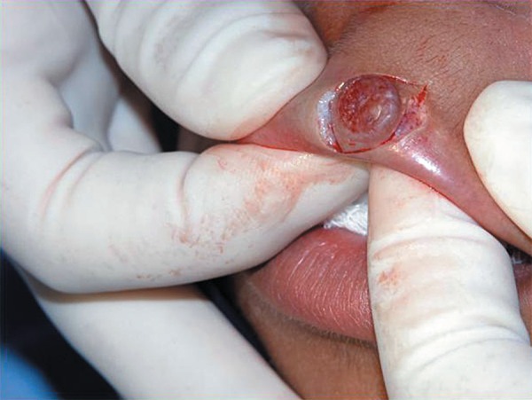
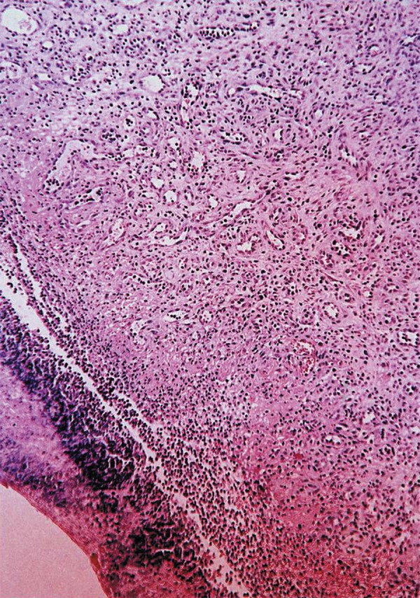
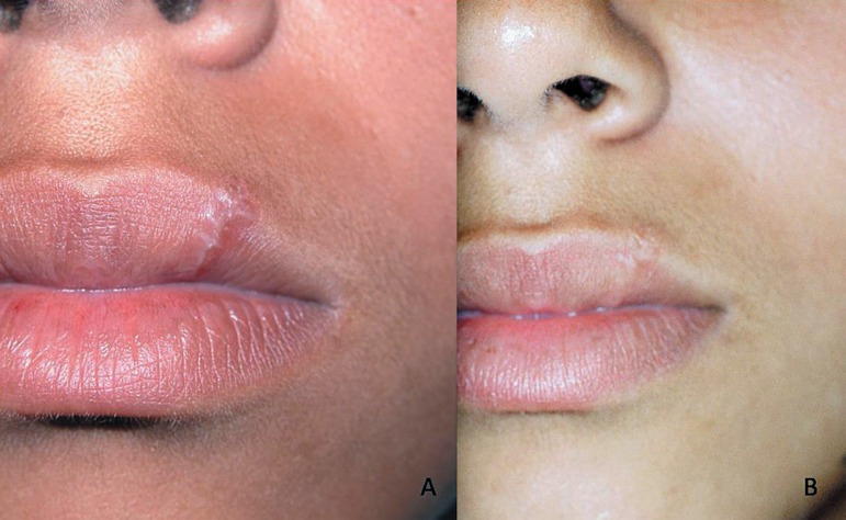
Pyogenic granuloma (PG) is a benign non-neoplastic mucocutaneous lesion. It is a reactional response to constant minor trauma and might be related to hormonal changes. In the mouth, PG is manifested as a sessile or pedunculated, resilient, erythematous, exophytic and painful papule or nodule with a smooth or lobulated surface that bleeds easily. PG preferentially affects the gingiva, but may also occur on the lips, tongue, oral mucosa and palate. The most common treatment is surgical excision. This paper describes a mucocutaneous PG on the upper lip, analyzing the clinical characteristics and discussing the features that distinguish this lesion from other similar oral mucosa lesions. The diagnosis of oral lesions is complex and leads the dentist to consider distinct lesions with different diagnostic methods. This case report with a 4 year-follow-up calls the attention to the uncommon mucocutaneous labial location of PG and to the fact that surgical excision is the safest method for diagnosis and treatment of PG of the lip, even when involving the mucosa and skin.
Figures
References
-
- Al-Khateeb T, Ababneh K. Oral pyogenic granuloma in Jordanians: a retrospective analysis of 108 cases. J Oral Maxillofac Surg. 2003;61:1285–1288. - PubMed
-
- Damm DD, Fantasia JE. Elevated and ulcerated nodule of lip. Pyogenic granuloma. Gen Dent. 2002;50:466–468. - PubMed
-
- Galeckas KJ, Uebelhoer NS. Successful treatment of pyogenic granuloma using a 1,064-nm laser followed by glycerin sclerotherapy. Dermatol Surg. 2009;35:530–534. - PubMed
-
- Graham RM. Pyogenic granuloma: an unusual presentation. Dent Update. 1996;23:240–241. - PubMed
-
- Ichimiya M, Yoshikawa Y, Hamamoto Y, Muto M. Successful treatment of pyogenic granuloma with injection of absolute ethanol. J Dermatol. 2004;31:342–344. - PubMed
Publication types
MeSH terms
LinkOut - more resources
Full Text Sources
