. 2011 Mar;43(3):478-84.
doi: 10.1007/s12031-010-9470-6. Epub 2010 Nov 18.

Astrocytes modulate distribution and neuronal signaling of leptin in the hypothalamus of obese A vy mice

Affiliations

Astrocytes modulate distribution and neuronal signaling of leptin in the hypothalamus of obese A vy mice

Weihong Pan et al. J Mol Neurosci. 2011 Mar.

Abstract

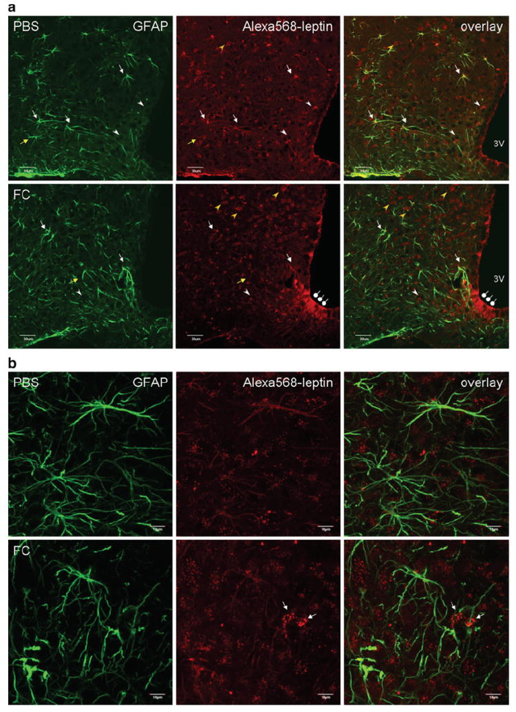
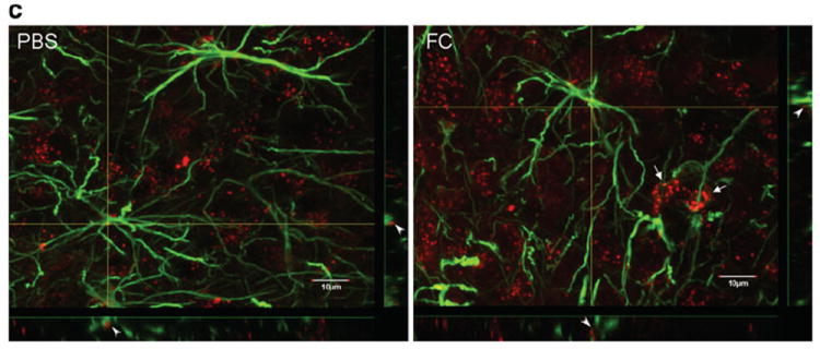
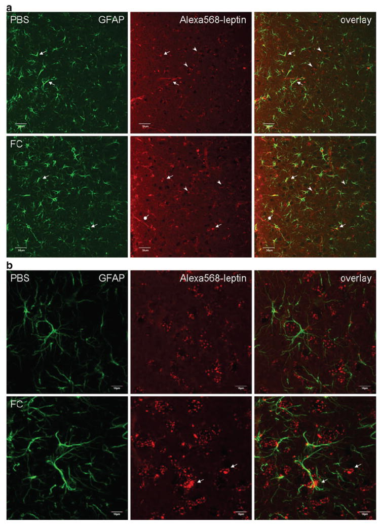
We tested the hypothesis that astrocytic activity modulates neuronal uptake and signaling of leptin in the adult-onset obese agouti viable yellow (A vy) mouse. In the immunohistochemical study, A vy mice were pretreated with the astrocyte metabolic inhibitor fluorocitrate or phosphate-buffered saline (PBS) vehicle intracerebroventricularly (icv) followed 1 h later by Alexa568-leptin. Confocal microscopy showed that fluorocitrate pretreatment reduced astrocytic uptake of Alexa568-leptin 30 min after icv while increasing neuronal uptake in the arcuate nucleus and dorsomedial hypothalamus. Fluorocitrate also induced mild astrogliosis and moderately increased pSTAT3 immunopositive neurons in response to Alexa568-leptin in the dorsomedial hypothalamus. In the Western blotting study, A vy mice were pretreated with either PBS or fluorocitrate, and received PBS or leptin 1 h later followed by determination of pSTAT3 and GFAP expression an additional 30 min afterward. The results show that fluorocitrate induced a mild pSTAT3 activation but attenuated leptin-induced pSTAT3 activation and decreased GFAP expression independently of leptin treatment. We conclude that inhibition of astrocytic activity resulted in enhanced neuronal leptin uptake and signaling. This suggests opposite roles of astrocytes and neurons in leptin's actions in the A vy mouse with adult-onset obesity.


Conflict of interest statement

Disclosures/Conflicts of Interest The authors have no conflict of interest and nothing to disclose.

Figures

Fig. 1
Fig. 1
Distribution of Alexa568-leptin (red) in the arcuate nucleus 30 min after icv administration, and partial colocalization with GFAP immunopositive cells (green). FC treatment increased the neuronal distribution of Alexa568-leptin. a In a representative brain section from a mouse receiving the control PBS, Alexa568-leptin is present in GFAP-positive cells (white arrows) and GFAP-negative cells that show neuronal morphology (yellow arrows). The Alexa dye shows either a vesicular distribution (white arrowheads) or a homogeneous pattern in the cytoplasm (yellow arrowheads). In a representative brain section from an FC pretreated mouse, there is a reduction of Alexa568 intensity in the GFAP-positive cells but an increase of Alexa568 (+) neurons that show a diffuse cytoplasmic staining pattern. FC pretreatment also induced subtle gliotic morphology in the astrocytes and increased the permeation of Alexa568-leptin along β1 tanycytes (oval arrows). 3V ventral third ventricle. Scale bars 30 μm. b From higher magnification confocal z-stacking sections in an enlarged area in the arcuate nucleus, Alexa568-leptin is seen both in astrocytes (homogeneous, corresponding to GFAP immmoreactivity) and neurons (vesicular pattern of cytoplasmic fluorescence). In the FC pretreated group, there is a reduction of Alexa568-leptin intensity in GFAP (+) cells, an increase in neurons adjacent to the GFAP (+) cells (arrows), and a ruffled morphology of GFAP (+) processes in contrast to the more extended filaments in the PBS pretreated group. c Overlay images with z-lines showing the presence of Alexa568-leptin in GFAP (+) cells (arrowheads) in the PBS pretreated group and lack of colocalization in the FC pretreated group. Scale bars 10 μm
Fig. 1
Fig. 1
Distribution of Alexa568-leptin (red) in the arcuate nucleus 30 min after icv administration, and partial colocalization with GFAP immunopositive cells (green). FC treatment increased the neuronal distribution of Alexa568-leptin. a In a representative brain section from a mouse receiving the control PBS, Alexa568-leptin is present in GFAP-positive cells (white arrows) and GFAP-negative cells that show neuronal morphology (yellow arrows). The Alexa dye shows either a vesicular distribution (white arrowheads) or a homogeneous pattern in the cytoplasm (yellow arrowheads). In a representative brain section from an FC pretreated mouse, there is a reduction of Alexa568 intensity in the GFAP-positive cells but an increase of Alexa568 (+) neurons that show a diffuse cytoplasmic staining pattern. FC pretreatment also induced subtle gliotic morphology in the astrocytes and increased the permeation of Alexa568-leptin along β1 tanycytes (oval arrows). 3V ventral third ventricle. Scale bars 30 μm. b From higher magnification confocal z-stacking sections in an enlarged area in the arcuate nucleus, Alexa568-leptin is seen both in astrocytes (homogeneous, corresponding to GFAP immmoreactivity) and neurons (vesicular pattern of cytoplasmic fluorescence). In the FC pretreated group, there is a reduction of Alexa568-leptin intensity in GFAP (+) cells, an increase in neurons adjacent to the GFAP (+) cells (arrows), and a ruffled morphology of GFAP (+) processes in contrast to the more extended filaments in the PBS pretreated group. c Overlay images with z-lines showing the presence of Alexa568-leptin in GFAP (+) cells (arrowheads) in the PBS pretreated group and lack of colocalization in the FC pretreated group. Scale bars 10 μm
Fig. 2
Fig. 2
Distribution of Alexa568-leptin (red) in the DMH 30 min after icv administration, its partial colocalization with GFAP immunopositive astrocytic cells (green), and activation of STAT3. FC treatment increased the neuronal distribution of Alexa568-leptin and the number of pSTAT3 immunopositive cells. a In the PBS pretreated group, there are fewer GFAP cells that internalized Alexa568-leptin (arrows) in the DMH than those in the arcuate nucleus. In neurons, the endocytosed Alexa568-leptin is present in perinuclear regions (arrowheads). In the FC pretreated group, tanycytes at the left border of the image are also Alexa568-leptin positive (oval arrow). The third ventricle is at the left border of each image. Scale bars 30 μm. b In higher magnification images with z-stacking, Alexa568-leptin has a vesicular distribution in the cytoplasm of neurons. There is an increase of Alexa568-leptin fluorescent intensity in neurons adjacent to the GFAP astrocytes in the FC pretreatment group (arrows). FC also induced an altered morphology, as the GFAP (+) processes appear more compact. Scale bars 10 μm. c Regional difference and effects of FC on pSTAT3 distribution in mice 30 min after Alexa568-leptin icv. There are more pSTAT3 (+) cells in the arcuate nucleus (ARC) than DMH. FC treatment increased the amount of pSTAT3 (+) cells in the DMH whereas that in the ARC did not show apparent change. Scale bars 50 μm. d pSTAT3 activation by leptin was seen by WB and reduced by FC pretreatment. In mice receiving FC, leptin did not show an additional effect on the level of pSTAT3 activation but GFAP expression was reduced in comparison with the internal control β-actin
Fig. 2
Fig. 2
Distribution of Alexa568-leptin (red) in the DMH 30 min after icv administration, its partial colocalization with GFAP immunopositive astrocytic cells (green), and activation of STAT3. FC treatment increased the neuronal distribution of Alexa568-leptin and the number of pSTAT3 immunopositive cells. a In the PBS pretreated group, there are fewer GFAP cells that internalized Alexa568-leptin (arrows) in the DMH than those in the arcuate nucleus. In neurons, the endocytosed Alexa568-leptin is present in perinuclear regions (arrowheads). In the FC pretreated group, tanycytes at the left border of the image are also Alexa568-leptin positive (oval arrow). The third ventricle is at the left border of each image. Scale bars 30 μm. b In higher magnification images with z-stacking, Alexa568-leptin has a vesicular distribution in the cytoplasm of neurons. There is an increase of Alexa568-leptin fluorescent intensity in neurons adjacent to the GFAP astrocytes in the FC pretreatment group (arrows). FC also induced an altered morphology, as the GFAP (+) processes appear more compact. Scale bars 10 μm. c Regional difference and effects of FC on pSTAT3 distribution in mice 30 min after Alexa568-leptin icv. There are more pSTAT3 (+) cells in the arcuate nucleus (ARC) than DMH. FC treatment increased the amount of pSTAT3 (+) cells in the DMH whereas that in the ARC did not show apparent change. Scale bars 50 μm. d pSTAT3 activation by leptin was seen by WB and reduced by FC pretreatment. In mice receiving FC, leptin did not show an additional effect on the level of pSTAT3 activation but GFAP expression was reduced in comparison with the internal control β-actin

Similar articles

Cited by

References

    1. Banks WA, Kastin AJ, Huang W, Jaspan JB, Maness LM. Leptin enters the brain by a saturable system independent of insulin. Peptides. 1996;17:305–311. - PubMed
    1. Blazquez JL, Rodriguez EM. The design of barriers in the hypothalamus allows the median eminence and the arcuate nucleus to enjoy private milieus: the former opens to the portal blood and the latter to the cerebrospinal fluid. Peptides. 2010;31:757–776. - PubMed
    1. Clarke DD. Fluoroacetate and fluorocitrate: mechanism of action. Neurochem Res. 1991;16:1055–1058. - PubMed
    1. He Y, Kastin AJ, Hsuchou H, Pan W. The Cdk5/p35 kinases modulate leptin-induced STAT3 signaling. J Mol Neurosci. 2009;39:49–58. - PMC - PubMed
    1. Hsuchou H, He Y, Kastin AJ, Tu H, Markadakis EN, Rogers RC, Fossier PB, Pan W. Obesity induces functional astrocytic leptin receptors in hypothalamus. Brain. 2009;132:889–902. - PMC - PubMed

Publication types

LinkOut - more resources