. 2011 Aug;68(16):2769-84.
doi: 10.1007/s00018-010-0591-4. Epub 2010 Nov 18.

Calcium-induced cleavage of DNA topoisomerase I involves the cytoplasmic-nuclear shuttling of calpain 2

Affiliations

Calcium-induced cleavage of DNA topoisomerase I involves the cytoplasmic-nuclear shuttling of calpain 2

Shang-Min Chou et al. Cell Mol Life Sci. 2011 Aug.

Abstract

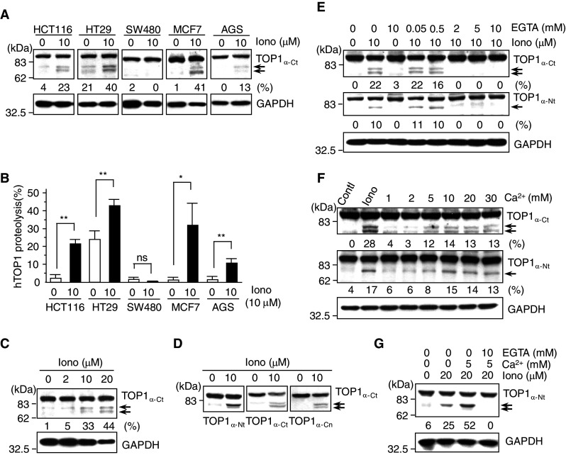
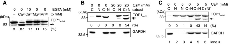
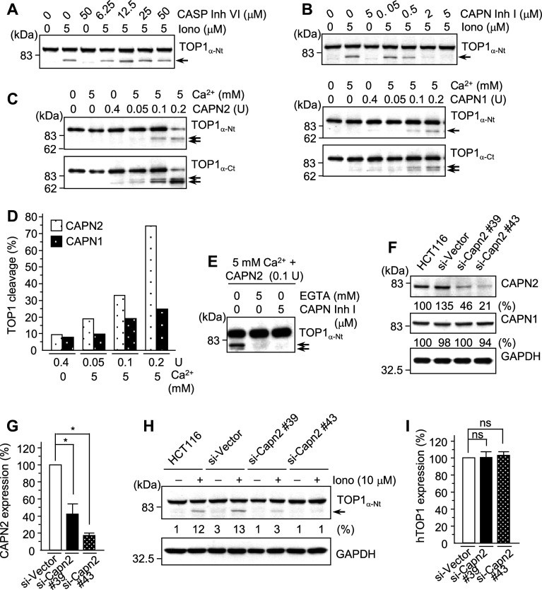
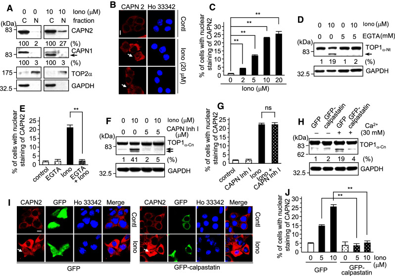
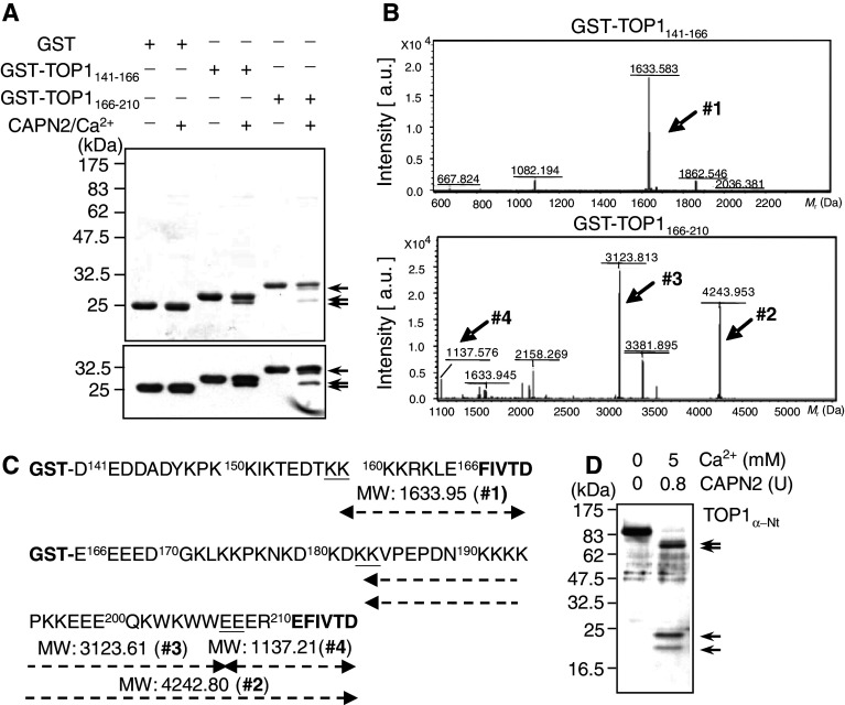
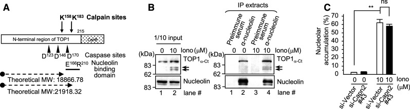
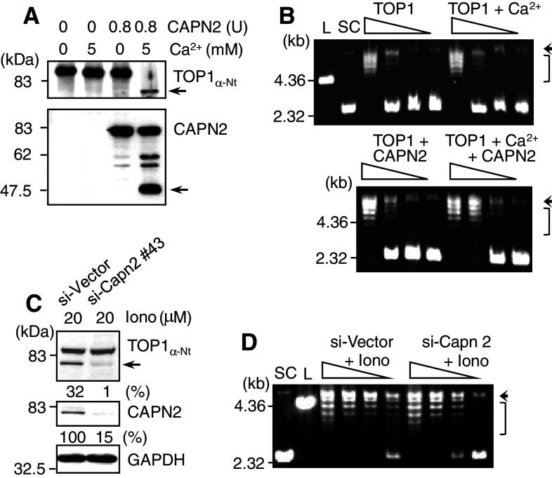
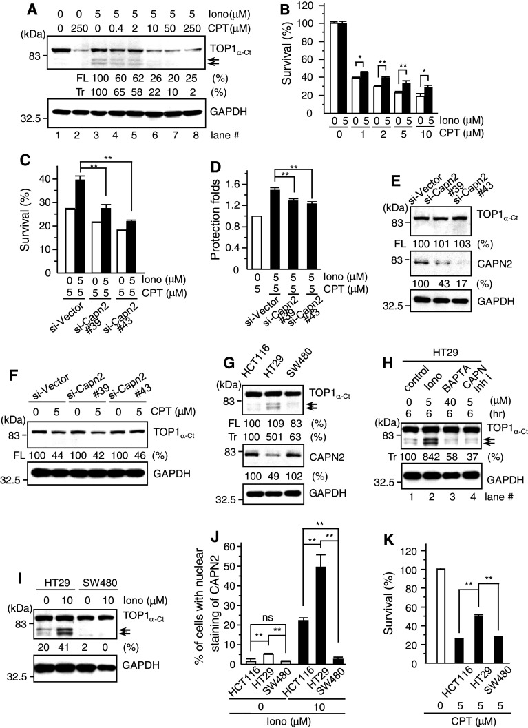
Important to the function of calpains is temporal and spatial regulation of their proteolytic activity. Here, we demonstrate that cytoplasm-resident calpain 2 cleaves human nuclear topoisomerase I (hTOP1) via Ca(2+)-activated proteolysis and nucleoplasmic shuttling of proteases. This proteolysis of hTOP1 was induced by either ionomycin-caused Ca(2+) influx or addition of Ca(2+) in cellular extracts. Ca(2+) failed to induce hTOP1 proteolysis in calpain 2-knockdown cells. Moreover, calpain 2 cleaved hTOP1 in vitro. Furthermore, calpain 2 entered the nucleus upon Ca(2+) influx, and calpastatin interfered with this process. Calpain 2 cleavage sites were mapped at K(158) and K(183) of hTOP1. Calpain 2-truncated hTOP1 exhibited greater relaxation activity but remained able to interact with nucleolin and to form cleavable complexes. Interestingly, calpain 2 appears to be involved in ionomycin-induced protection from camptothecin-induced cytotoxicity. Thus, our data suggest that nucleocytoplasmic shuttling may serve as a novel type of regulation for calpain 2-mediated nuclear proteolysis.


Figures

Fig. 1
Fig. 1
Ionomycin treatment induced rapid, but limited proteolysis of human DNA topoisomerase I (hTOP1) in cells. a Cellular exposure to ionomycin resulted in the cleavage of hTOP1 proteins in various cell lines. HCT116, HT29, SW480 colorectal, MCF7 breast and AGS gastric cancer cells were treated with or without ionomycin (Iono, 10 μM) for 15 min, harvested and lysed. Equal amounts of different cell lysates were subjected to immunoblotting analysis with antibodies against the C-terminus of TOP1 (TOP1α-Ct) and GAPDH (as the loading control) and proteolysis results were quantified (number of replicates, n = 3) (b). c Ionomycin-induced hTOP1 proteolysis is dose-dependent. d The proteolysis of hTOP1 induced by ionomycin is likely caused by the protease-mediated cleavage at the N′-terminus. HCT116 cells were treated with ionomycin (doses as indicated) and the integrity of proteins was analyzed as described in a. Antibodies against the central domain, N-terminus, and C-terminus of hTOP1 (named TOP1α-Cn, TOP1α-Nt, and TOP1α-Ct, respectively) were used for the epitope-mapping of potential proteolytic sites on hTOP1. e Ca2+ chelation by EGTA prevented hTOP1 proteolysis induced by ionomycin. HCT116 cells were pre-treated with EGTA for 30 min and then underwent co-treatment with ionomycin for 15 min. f The hTOP1 proteolysis was also observed with extracellular addition of Ca2+ to the culture media. Different concentrations of Ca2+ (as indicated on the top of figure) were add directly into the media and HCT116 cells were further incubated for 15 min. g The addition of extracellular Ca2+ also greatly stimulated ionomycin-induced proteolysis of hTOP1. HCT116 cells were treated with different combinations of 5-mM Ca2+, 10-mM EGTA (pre-treatment for 30 min) and 20-μM ionomycin for 15 min. Cells were lysed, and immunoblotting analyses were carried out as described above. Arrows indicate the truncated hTOP1 fragments. **p < 0.01; * p < 0.05; ns, statistically non-significant
Fig. 2
Fig. 2
In vitro proteolytic cleavage of nuclear hTOP1 by Ca2+-activated cytosolic proteases. a Ca2+, but not Mg2+ or Mn2+, effectively activated hTOP1 proteolysis in HCT116 cell extracts. The in vitro protease activation assay using total extracts was performed in the absence or presence of different divalent cations (5 mM) as described in “Materials and methods”. b The fraction and reconstitution experiments revealed that cytosolic extract contains the Ca2+-activated proteolytic activity on hTOP1. c The intact nuclear membrane partially blocked the Ca2+-activated cleavage of hTOP1. The Ca2+-activated hTOP1 proteolytic reaction was reconstituted by various combinations of cytosolic extract (C), nuclear extract (N) and isolated nuclei (Ni′) as described in “Materials and methods” (n = 2). Ca2+ (5 mM) was added to activate the protease(s). After incubation at room temperature for 10 min, the mixtures were subjected to immunoblotting analysis with hTOP1α-Nt antibodies
Fig. 3
Fig. 3
Calpain 2 is the main protease responsible for the Ca2+-activated proteolysis of hTOP1. a Ionomycin could still effectively induce hTOP1 proteolysis in the presence of caspase inhibitor. b Ionomycin-induced hTOP1 proteolysis was greatly reduced when calpain proteases were inhibited. Different concentrations (μM, as indicated on the top of corresponding panels) of caspase inhibitor VI (CASP Inh VI) and calpain inhibitor I (CAPN Inh I) were added for 30 min before ionomycin treatment (5 μM for 15 min). c, d The proteolysis of purified hTOP1 by recombinant calpain 1 (CAPN1) and 2 (CAPN2) in vitro. Purified calpains (human CAPN1 or rat CAPN2, at indicated units) with or without Ca2+ (5 mM) were mixed with recombinant hTOP1 (0.05 μg) and incubated for 10 min at room temperature; then, reactions were stopped using sample buffer. The quantitative data has been plotted in (d). e Both Ca2+-chelating EGTA and calpain inhibitor I efficiently blocked in vitro proteolysis of hTOP1 mediated by the Ca2+-activated recombinant calpain 2. f, g The expression of calpain 2 was specifically knocked-down using a RNA interference (RNAi) approach. The knockdown efficiencies in HCT116 cells of lentiviral particles expressing different shRNAs (#39 and #43) were quantified (n = 3) (g). h Ca2+-mediated hTOP1 proteolysis was diminished in the calpain 2-deficient cells. The lentivirus-mediated RNAi approach was utilized to specifically knockdown the expression of calpain 2 in HCT116 cells. After proper selection, pooled clones of calpain 2-deficient cell lines were established. The knockdown efficiency and specificity values for these two clones (HCT116 si-Capn2 #39 and #43) are shown in (f, g). Ionomycin-induced hTOP1 proteolysis was then performed with two knockdown clones and HCT116 si-Vector cells as described above. i The expression levels of hTOP1 are not altered in two si-Capn2 cell lines (n = 3). * p < 0.05; ns, statistically non-significant
Fig. 4
Fig. 4
Cytosolic calpain 2 entered the nucleus and cleaved hTOP1 after Ca2+ influx. a, b Ionomycin treatment shifted a fraction of cytoplasmic calpain 2 into the nucleus as revealed by the fractionation assay (a) and the immunofluorescent analysis (IFA) using a confocal microscope (b). Fractionation experiments were performed with HCT116 cells treated with or without ionomycin (10 μM, 15 min). Cytosolic and nuclear extracts were then analyzed by the immunoblotting analysis using anti-calpain 1 and 2 antibodies. GAPDH and hTOP2α were used as cytosolic and nuclear markers, respectively. For IFA, cells were first seeded onto cover-slips for 24 h before ionomycin treatment (20 μM for 15 min). Control and treated cells were subjected to IFA using a confocal microscope as described in “Materials and methods”. Nuclear location was indicated by Hoechst 33342 staining. Bar 8 μM. c Quantitative results for the dosage dependence of ionomycin-induced nuclear entry of calpain 2 (n = 3). d, e Chelation of extracellular Ca2+ by EGTA effectively abolished both ionomycin-induced proteolysis of hTOP1 (d) and nuclear entry of calpain 2 (n = 3) (e). f, g Inhibition of protease activity by calpain inhibitor I (CAPN Inh I) affected only the ionomycin-induced hTOP1 proteolysis (f), but not the nuclear entry of calpain 2 (n = 3) (g). Ectopic expression of calpastatin reduced both Ca2+-activated proteolysis of hTOP1 (h) and ionomycin-induced nuclear entry of calpain 2 (i). HCT116 cells were transfected with either a control vector or the GFP-calpastatin fusion-expressing construct. After 48 h, the GFP fluorescence assay was used to examine the transfection efficiencies (~30–40%). j Quantitative analysis of the effect of calpastatin expression on ionomycin-induced nuclear entry of calpain 2 (n = 3). Columns represent percentages of nuclei containing calpain 2 in GFP-positive cells. White arrow heads indicated for the cells with nuclear staining of calpain 2. **p < 0.01; ns, statistically non-significant
Fig. 5
Fig. 5
Identification of the calpain 2-dependent cleavage sites in hTOP1 by mass spectrometry. a Purified hTOP1 fragments were cleaved by calpain 2 in vitro. b MALDI-TOF/MS analysis results for the calpain 2-cleaved products of GST-tagged hTOP1 fragments. Two GST-fused hTOP1 fragments, GST-TOP1141–166 and GST-TOP1166–210 proteins (with amino acid 141–166 and 166–210 of hTOP1, respectively), were expressed and purified from bacteria. The calpain 2 proteolytic reactions were performed with these two GST-fused tagged hTOP1 fragments and the reaction mixtures were then subjected to SDS-PAGE separation (a, upper panel; stained with Coomassie Blue), immunoblotting analysis with anti-GST antibodies (a, lower panel) and MALDI-TOF/MS analysis (b). The calpain 2-truncated products, the intact and truncated GST-containing hTOP1 fragments in the reaction mixtures were loaded into Microcon YM-10 columns. The flow-through parts were collected for molecular weight determination by MALDI-TOF mass spectrometry analysis. The molecular masses determined for the calpain 2-cleaved hTOP1 short fragments in the flow-through are shown underlined on the top of the peaks (b, units = Da). c Schematic mapping containing hTOP1 amino acid sequences of GST-TOP1141–166 and GST-TOP1166–210 peptides and the theoretical molecular masses for the calpain 2-cleaved fragments (arrows #1–4 shown in b). The non-hTOP1-derived amino acids FIVTD and EFIVTD (in bold) are encoded from the backbone of the pGEX-1λ-T vector after cloning, and the two calpain 2 cleavage sites in hTOP1 are underlined. d Calpain 2 cleaved hTOP1at two sites to produce in four fragments. After the in vitro calpain 2 cleavage of hTOP1, the reaction mixtures were subjected to immunoblotting analysis. The arrow heads indicate the cleaved hTOP1 fragments. MW, molecular weight; a.u., arbitrary units
Fig. 6
Fig. 6
Ionomycin treatment enhanced the nucleolar distribution of hTOP1, and calpain 2-mediated proteolysis of hTOP1 did not disrupt its interaction with nucleolin. a Diagram indicating two calpain 2 cleavage sites, three caspase cleavage sites and the nucleolin-interacting domain identified on the N-terminus of hTOP1. The two calpain 2 cleavage sites (K158 and K183) mapped in this study and three caspase cleavage sites (D123, D146 and D170) are indicated by the arrows and arrowheads, respectively. The nucleolin-binding domain (E166 to R210) of hTOP1 is also presented. b Full-length hTOP1 and calpain 2-truncated hTOP1tr both interacted with nucleolin. Ionomycin-treated or control HCT116 cell lysates were subjected to the immune-precipitation (IP) assay as described using antibodies against nucleolin. c Nucleolar accumulation of hTOP1 is regulated by Ca2+ influx, but independent of calpain 2. HCT116 si-Vector and si-Capn2 #43 cells were seeded on cover-slips for 24 h before exposure to 10 μM ionomycin for 15 min, and hTOP1 localization was examined by immunofluorescence analysis as described. MW, molecular weight; **p < 0.01; ns, statistically non-significant
Fig. 7
Fig. 7
hTOP1 proteins truncated by calpain 2 exhibit greater relaxation activity than full-length hTOP1. a Addition of Ca2+ caused proteolysis of both calpain 2 and hTOP1 in vitro. b Calpain 2-mediated cleavage enhanced the relaxation activity of hTOP1-containing mixtures. Equal amounts of purified hTOP1 proteins were mixed with Ca2+ and/or calpain 2 and the reaction mixtures were incubated for 10 min at room temperature. The relative relaxation activities of reaction mixtures were determined with a twofold serial dilution as described in “Materials and methods”. c, d Ionomycin treatment to calpain 2-proficient cells increased both the proteolysis of hTOP1 and relaxation activity of cellular extracts. HCT116 si-Capn2 #43 and si-Vector cells were treated with 20-μM ionomycin for 15 min, lysed and collected for the immunoblotting analysis (c) and relaxation assay (d). Arrows, truncated hTOP1 or calpain 2 fragments (a and c); arrow heads, nick-from DNA (b and d); L, linearized DNA; SC, supercoiled DNA; Iono, ionomycin; Bracket, DNA topoisomers
Fig. 8
Fig. 8
Ca2+-activated calpain 2 cleavage compromised TOP1-mediated cell killing. Cellular exposure to TOP1-targeting camptothecin (CPT, 30 min) resulted in the formation of both the full-length hTOP1 cleavable complex (hTOP1 cc) and truncated hTOP1trcc in HCT116 cells (a). The formation of CPT-induced hTOP1 cc and hTOP1trcc in the trapping assay is indicated by the disappearance of hTOP1. The levels of full-length (FL) and truncated (Tr) hTOP1 proteins in the ionomycin-treated sample were both taken as 100%. b Ionomycin treatment protected cells from the cytotoxic action of CPT. HCT116 cells pre-treated with or without ionomycin (15 min, concentrations as indicated) were co-incubated with CPT (conc. as indicated in figures) for 30 min. The trapping assay and colony formation assay were performed as described in “Materials and methods” to quantify CPT-induced hTOP1 cc formation (a) and cell killing (n = 3) (b). c, d Calpain 2 is involved in the ionomycin-induced protection from CPT cytotoxicity. HCT116 si-Vector, si-Capn2 #39 or #43 cells were exposed to ionomycin (5 μM) and CPT (5 μM) and cell survival was determined in three independent experiments (n = 3). The relative protective effects of ionomycin against CPT-induced cell killing in different cell lines were further quantified using the ratio of survival rates in the presence and absence of ionomycin and represent as “protection folds” in (d). e The hTOP1 expression level was not affected by the knockdown of calpain 2 in HCT116 cells. f CPT induced almost identical formation of hTOP1 cc in HCT116 si-Vector and si-Capn 2 cells. Cells were treated with 5-μM CPT for 30 min and the formation of hTOP1 cc was assayed as described in Fig. 8a (n = 3). g HT29 cells exhibited the highest basal level of hTOP1 proteolysis among the three colorectal cancer cell lines. h Both the Ca2+ chelator BAPTA and calpain inhibitor I effectively reduced the endogenous level of hTOP1 proteolysis. HT29 cells incubated with 5-μM ionomycin, 40-μM BAPTA or 5-μM calpain inhibitor I (CAPN Inh I) for 6 h and hTOP1 proteins were assayed as described above. The endogenous level of truncated (Tr) hTOP1 in HT29 cells (lane 1) was taken as 100%. Ionomycin treatment induced differential proteolysis of hTOP1 (i) and nuclear entry of calpain 2 (j) in three different colorectal cell lines. k The difference in sensitivity to the CPT-mediated cell killing correlates with a differential hTOP1 proteolysis ability in the three colorectal cancer cell lines. Experiments performed with three colorectal cancer cells, HT29, HCT116, and SW480, were as described above (n = 3). FL, full-length hTOP1; Tr, calpain 2-truncated hTOP1 fragments; **p < 0.01; ns, statistically non-significant

Similar articles

Cited by

References

    1. Bartoli M, Richard I. Calpains in muscle wasting. Int J Biochem Cell Biol. 2005;37:2115–2133. doi: 10.1016/j.biocel.2004.12.012. - DOI - PubMed
    1. Tsuji T, Shimohama S, Kimura J, Shimizu K. m-Calpain (calcium-activated neutral proteinase) in Alzheimer’s disease brains. Neurosci Lett. 1998;248:109–112. doi: 10.1016/S0304-3940(98)00348-6. - DOI - PubMed
    1. Vanderklish PW, Bahr BA. The pathogenic activation of calpain: a marker and mediator of cellular toxicity and disease states. Int J Exp Pathol. 2000;81:323–339. doi: 10.1046/j.1365-2613.2000.00169.x. - DOI - PMC - PubMed
    1. Green KN, LaFerla FM. Linking calcium to Abeta and Alzheimer’s disease. Neuron. 2008;59:190–194. doi: 10.1016/j.neuron.2008.07.013. - DOI - PubMed
    1. Dargelos E, Brule C, Combaret L, Hadj-Sassi A, Dulong S, Poussard S, Cottin P. Involvement of the calcium-dependent proteolytic system in skeletal muscle aging. Exp Gerontol. 2007;42:1088–1098. doi: 10.1016/j.exger.2007.08.009. - DOI - PubMed

Publication types

MeSH terms