

Selective improvement of tumor necrosis factor capture in a cytokine hemoadsorption device using immobilized anti-tumor necrosis factor
- PMID: 21086427
- PMCID: PMC3221482
- DOI: 10.1002/jbm.b.31748
Selective improvement of tumor necrosis factor capture in a cytokine hemoadsorption device using immobilized anti-tumor necrosis factor
Abstract
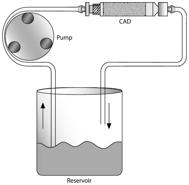
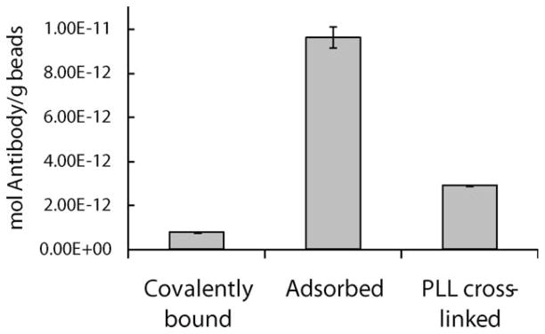
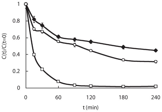
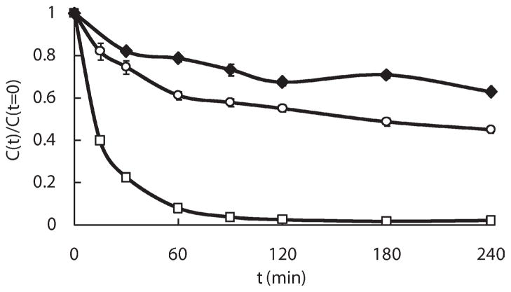
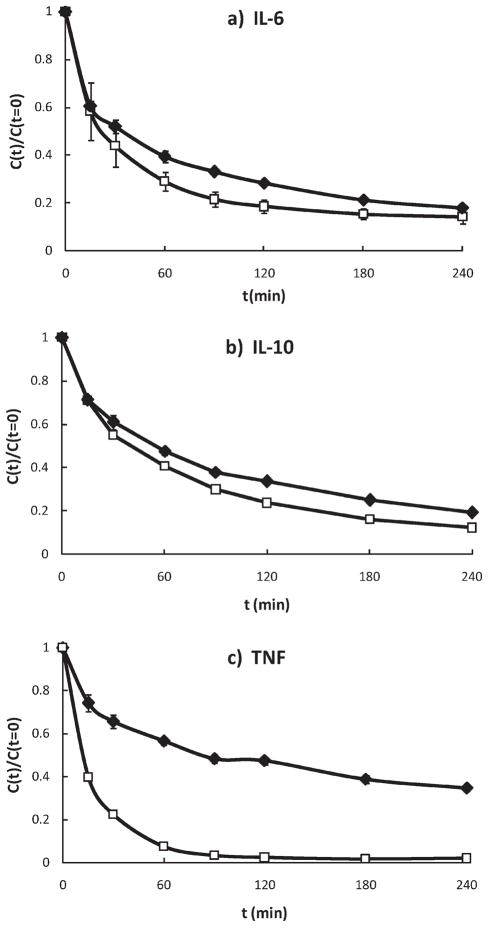
Sepsis is a harmful hyper-inflammatory state characterized by overproduction of cytokines. Removal of these cytokines using an extracorporeal device is a potential therapy for sepsis. We are developing a cytokine adsorption device (CAD) filled with porous polymer beads which efficiently depletes middle-molecular weight cytokines from a circulating solution. However, removal of one of our targeted cytokines, tumor necrosis factor (TNF), has been significantly lower than other smaller cytokines. We addressed this issue by incorporating anti-TNF antibodies on the outer surface of the beads. We demonstrated that covalent immobilization of anti-TNF increases overall TNF capture from 55% (using unmodified beads) to 69%. Passive adsorption increases TNF capture to over 99%. Beads containing adsorbed anti-TNF showed no significant loss in their ability to remove smaller cytokines, as tested using interleukin-6 (IL-6) and interleukin-10 (IL-10). We also detail a novel method for quantifying surface-bound ligand on a solid substrate. This assay enabled us to rapidly test several methods of antibody immobilization and their appropriate controls using dramatically fewer resources. These new adsorbed anti-TNF beads provide an additional level of control over a device which previously was restricted to nonspecific cytokine adsorption. This combined approach will continue to be optimized as more information becomes available about which cytokines play the most important role in sepsis.
© 2010 Wiley Periodicals, Inc.
Figures
Similar articles
-
A simple mathematical model of cytokine capture using a hemoadsorption device.Ann Biomed Eng. 2009 Jan;37(1):222-9. doi: 10.1007/s10439-008-9587-8. Epub 2008 Oct 24. Ann Biomed Eng. 2009. PMID: 18949559 Free PMC article.
-
Effects of hemoadsorption with a novel adsorbent on sepsis: in vivo and in vitro study.Blood Purif. 2015;39(1-3):239-245. doi: 10.1159/000381006. Epub 2015 Apr 1. Blood Purif. 2015. PMID: 25833160 Free PMC article.
-
Characterizing accelerated capture of deoligomerized TNF within hemoadsorption beads used to treat sepsis.J Biomed Mater Res B Appl Biomater. 2011 Jul;98(1):47-53. doi: 10.1002/jbm.b.31830. Epub 2011 Apr 18. J Biomed Mater Res B Appl Biomater. 2011. PMID: 21504054
-
Hemofiltration, adsorption, sieving and the challenge of sepsis therapy design.Crit Care. 2002 Oct;6(5):394-6. doi: 10.1186/cc1826. Epub 2002 Sep 4. Crit Care. 2002. PMID: 12398774 Free PMC article. Review.
-
Relevance of platelet-activating factor in inflammation and sepsis: mechanisms and kinetics of removal in extracorporeal treatments.Am J Kidney Dis. 1997 Nov;30(5 Suppl 4):S57-65. doi: 10.1016/s0272-6386(97)90543-6. Am J Kidney Dis. 1997. PMID: 9372980 Review.
Cited by
-
Adsorption of the inflammatory mediator high-mobility group box 1 by polymers with different charge and porosity.Biomed Res Int. 2014;2014:238160. doi: 10.1155/2014/238160. Epub 2014 Aug 27. Biomed Res Int. 2014. PMID: 25243124 Free PMC article.
-
Sepsis: mechanisms of bacterial injury to the patient.Scand J Trauma Resusc Emerg Med. 2019 Feb 14;27(1):19. doi: 10.1186/s13049-019-0596-4. Scand J Trauma Resusc Emerg Med. 2019. PMID: 30764843 Free PMC article. Review.
-
Antibody-modified conduits for highly selective cytokine elimination from blood.JCI Insight. 2018 Jul 12;3(13):e121133. doi: 10.1172/jci.insight.121133. JCI Insight. 2018. PMID: 29997301 Free PMC article.
-
A historical perspective on sepsis.Am J Pathol. 2012 Jul;181(1):2-7. doi: 10.1016/j.ajpath.2012.05.003. Epub 2012 May 26. Am J Pathol. 2012. PMID: 22642906 Free PMC article. Review.
References
-
- Angus DC, Linde-Zwirble WT, Lidicker J, Clermont G, Carcillo J, Pinsky MR. Epidemiology of severe sepsis in the United States: Analysis of incidence, outcome, and associated costs of care. Crit Care Med. 2001;29:1303–1310. - PubMed
-
- Nylen ES, Alarifi AA. Humoral markers of severity and prognosis of critical illness. Best Pract Res Clin Endocrinol Metab. 2001;15:553–573. - PubMed
-
- van der Poll T, van Deventer SJ. Cytokines and anticytokines in the pathogenesis of sepsis. Infect Dis Clin North Am. 1999;13:413–426. - PubMed
-
- Cross AS, Opal SM. A new paradigm for the treatment of sepsis: Is it time to consider combination therapy? Ann Intern Med. 2003;138:502–505. - PubMed
-
- Baue AE. Multiple organ failure, multiple organ dysfunction syndrome, and systemic inflammatory response syndrome. Why no magic bullets? Arch Surg. 1997;132:703–707. - PubMed
Publication types
MeSH terms
Substances
Grants and funding
LinkOut - more resources
Full Text Sources
Miscellaneous
