Review
. 2010 Dec;45(6):520-34.
doi: 10.3109/10409238.2010.513375.

Topoisomerases and site-specific recombinases: similarities in structure and mechanism

Affiliations
Review

Topoisomerases and site-specific recombinases: similarities in structure and mechanism

Wei Yang. Crit Rev Biochem Mol Biol. 2010 Dec.

Abstract

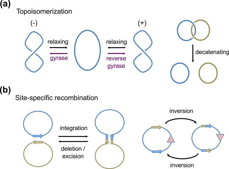
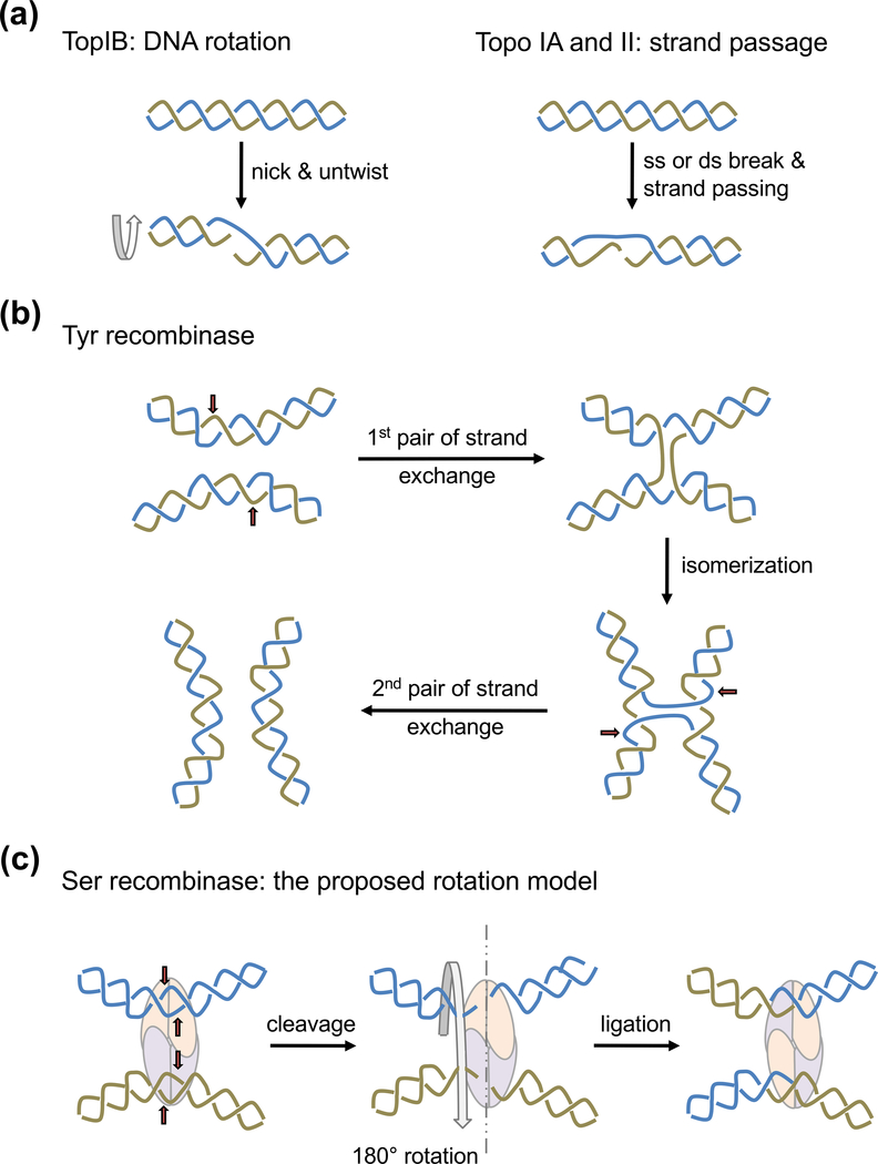
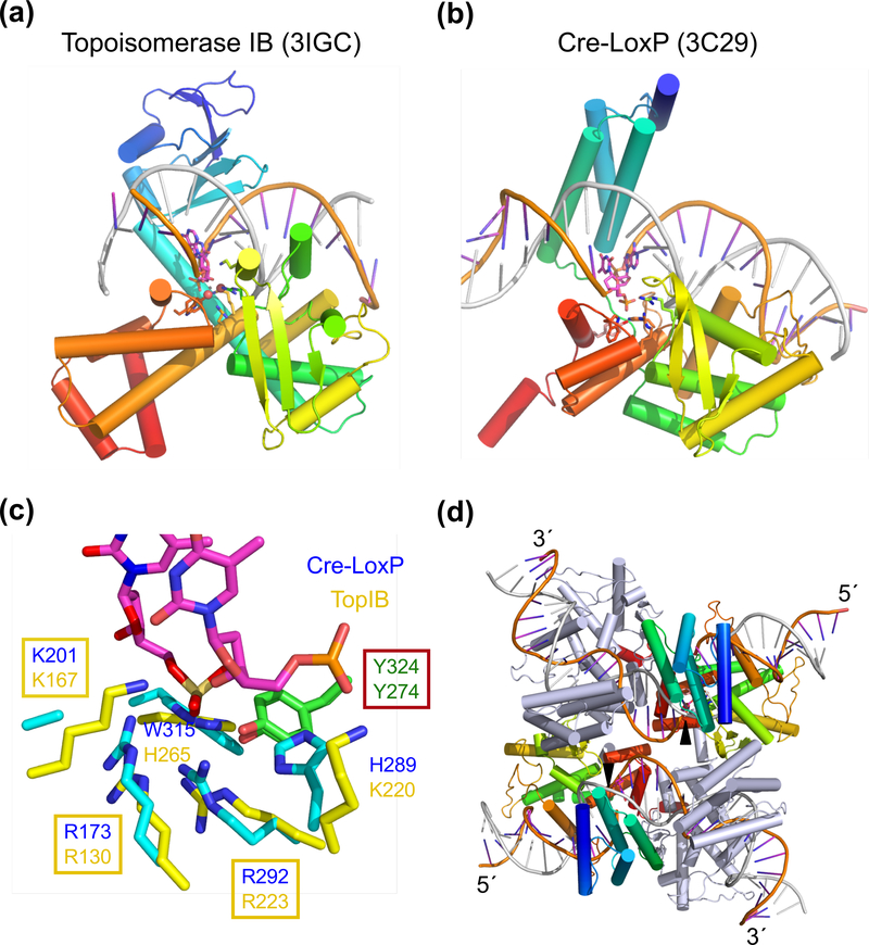
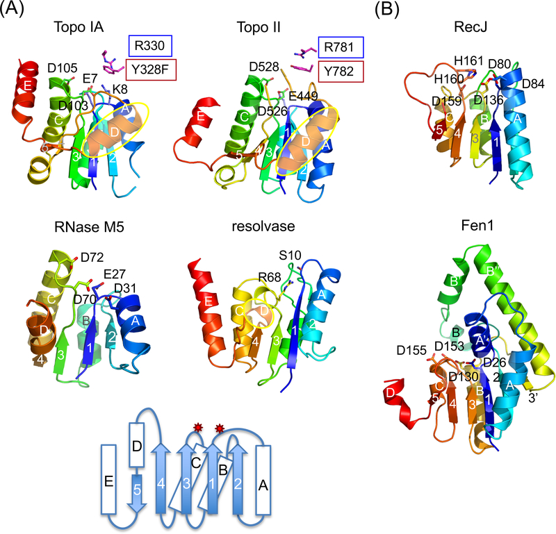
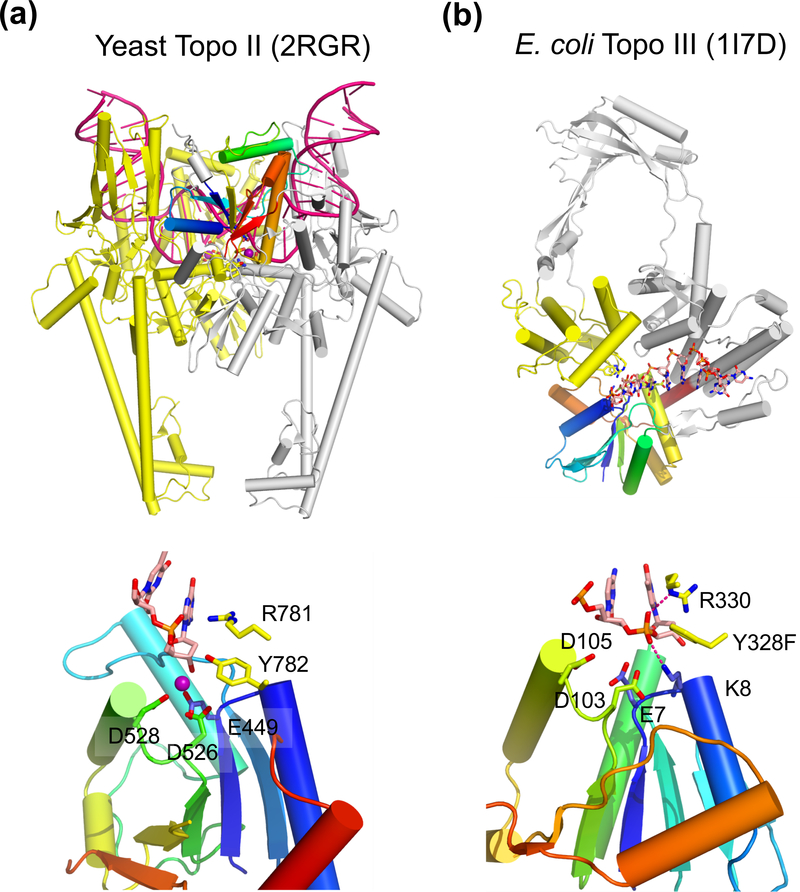
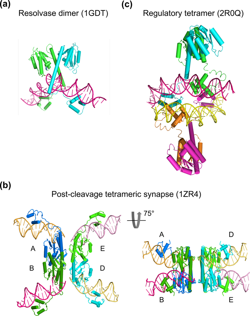
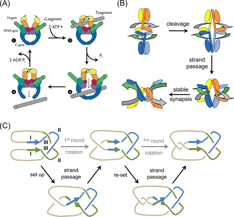
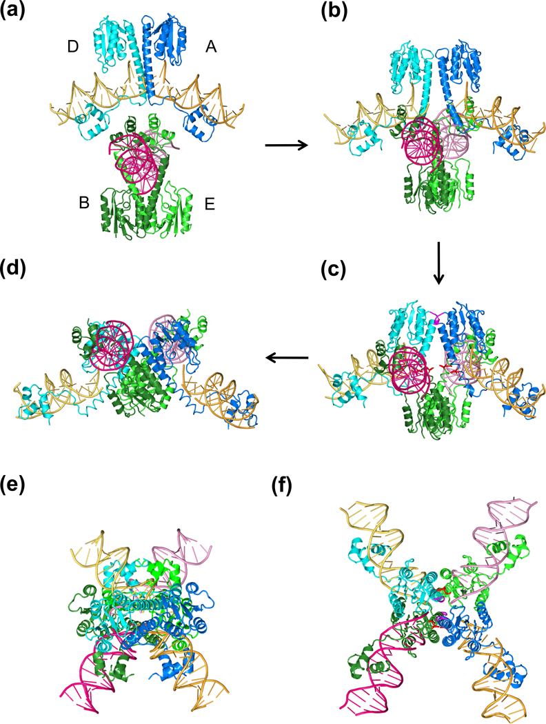
The processes of DNA topoisomerization and site-specific recombination are fundamentally similar: DNA cleavage by forming a phospho-protein covalent linkage, DNA topological rearrangement, and DNA ligation coupled with protein regeneration. Type IB DNA topoisomerases are structurally and mechanistically homologous to tyrosine recombinases. Both enzymes nick DNA double helices independent of metal ions, form 3'-phosphotyrosine intermediates, and rearrange the free 5' ends relative to the uncut strands by swiveling. In contrast, serine recombinases generate 5'-phospho-serine intermediates. A 180° relative rotation of the two halves of a 100 kDa terameric serine recombinase and DNA complex has been proposed as the mechanism of strand exchange. Here I propose an alternative mechanism. Interestingly, the catalytic domain of serine recombinases has structural similarity to the TOPRIM domain, conserved among all Type IA and Type II topoisomerases and responsible for metal binding and DNA cleavage. TOPRIM topoisomerases also cleave DNA to generate 5'-phosphate and 3'-OH groups. Based on the existing biochemical data and crystal structures of topoisomerase II and serine recombinases bound to pre- and post-cleavage DNA, I suggest a strand passage mechanism for DNA recombination by serine recombinases. This mechanism is reminiscent of DNA topoisomerization and does not require subunit rotation.


Conflict of interest statement

Declaration of Interest

The research was funded by the intramural research program of NIDDK, NIH.

Declaration of no interest

The author reports no conflicts of interest. The author alone is responsible for the content and writing of the paper.

Figures

Fig. 1
Fig. 1
Diagrams of topoisomerization and site-specific recombination. (a) DNA Topoisomerization by changing supercoils or decatenation of linked DNA circles. (b) Site-specific recombinases catalyze DNA integration, deletion (also called excision or resolution), and inversion. These reactions require two specific DNA sequences shown as arrows and negative supercols as the energy source. The color versions of all figure are available online.
Fig. 2
Fig. 2
Mechanisms of topoisomerization and recombination by YRs. (a) Two mechanisms are used for topoisomerization. Type IB topoisomerases (TopIB) relax (−) or (+) supercoils by nicking one strand and allowing DNA to untwist. Topo IA and Topo II can relax as well as make supercoils by breaking one strand or duplex and passing the other through it. The intertwined blue and beige color lines represent single strands in the case of Topo IA or double helices in the case of Topo II. (b) Tyrosine recombinases (YRs) catalyze DNA recombination in two steps. One pair of strands are cleaved, exchanged and rejoined in each step (beige and blue colored lines sequentially). The red arrows point the cleavage sites. A Holliday junction is formed and isomerized between the two steps. (c) Serine recombinases make concerted double strand breaks of two recombination sites. The proposed subunit rotation mechanism requires the left half of the cleaved DNA rotate 180° relative to the right half for strand exchange.
Fig. 3
Fig. 3
Structural comparison of TopIB and YRs. (a) The structure of smallpox viral topoisomerase complexed with DNA (PDB: 3IGC). (b) One subunit of Cre bound to one half of a loxP site (PDB: 3C29). In (a) and (b) each polypeptide chain is shown as blue (N-) to red (C-terminus) rainbow-colored ribbon diagrams. The DNA strand being nicked is colored orange and the complementary strand in silver. The active site residues are shown in sticks. (c) Superposition of the active sites of TopIB and YR. Both tyrosine nucleophiles are shown in green (carbon) and red (oxygen). Other conserved residues are shown in yellow (TopIB) and cyan (YR) with blue nitrogen. A trio of basic residues, RKR, is conserved among all TopIBs and YRs. Nearby His and Trp conserved among YRs are replaced, respectively, by Lys and His conserved in TopIBs. (d) The tetrameric Cre-loxP complex (PDB: 3C29). The view is roughly perpendicular to (b). The two Cre subunits poised to cleave DNA are shown in rainbow colors and the other two in grey. Once the orange-colored strands are cleaved (indicated by the black arrowheads), their 3´-ends are covalently linked to the Cre subunits, and the free 5´ ends reciprocally invade and rejoin with the neighboring DNAs thus forming a HJ. Isomerization of HJ activates the grey-colored Cre subunits to catalyze the exchange of silver-colored strands.
Fig. 4
Fig. 4
TOPRIM and SR catalytic domain are similar in the folding topology and active-site location. (a) TOPRIM domain of Topo IA (1I7D), II (2RGR) and RNase M5 (1T6T) are aligned with the catalytic domain of γδ resolvase (1GDT). Each is shown in rainbow-colored ribbon diagrams. Active site residues are highlighted as sticks. The D helices (in yellow ovals) in topoisomerases are longer than in resolvase. A topology diagram below summarizes all four examples. The catalytic residues are located on two loops indicated by the red stars. Three carboxylates (E, DxD) are conserved among all TOPRIM domains for metal-ion binding. M5 nuclease has an additional carboxylates (D31) and uses water as nucleophile. Topo IA has a catalytic Lys (K8) in the equivalent location. (b) Ribbon diagrams of the catalytic domain of RecJ (1IR6) and Fen1 (1EXN) in rainbow colors. The catalytic residues are shown and labeled. Inserted elements in Fen1 are labeled after the adjacent secondary structures with apostrophes (A’, B’, B” and 3’).
Fig. 5
Fig. 5
Structure of type II and type IA topoisomerase. (a) The overall structure of the dimeric yeast Topo II (type II) bound to a substrate DNA (G segment, PDB: 2RGR). One subunit is shown in silver with the TOPRIM domain in rainbow colors. The other subunit, which donates the tyrosine nucleophile to the rainbow-colored TOPRIM, is shown in yellow. The DNA substrate is severely bent and shown in dark pink. The N-terminal ATPase domain is absent in the crystal structure (not shown) and would be on top of DNA in this view. (b) E. coli Topo III (type IA, PDB: 1I7D) is monomeric. The TOPRIM is shown in rainbow colors. The tyrosine nucloephile is donated by the yellow domain. The rest of the protein is colored in silver, The ssDNA substrate is shown as multicolored sticks. The active site of Topo II and Topo III are shown below. In addition to the three carboxylates (E, DxD), the catalytically essential Arg and Tyr (nucleophile) are donated from the second subunit or domain (colored yellow) for catalysis. The Mg2+ found in Topo II is shown as a purple sphere.
Fig. 6
Fig. 6
Crystal structures of resolvase-DNA complexes. (a) The pre-cleavage resolvase dimer-site I DNA complex (PDB: 1GDT). The protein subunits are shown as cyan and green ribbon diagrams and DNA in pink tube-and-ladder. (b) Two views of a post-cleavage tetrameric resolvase complex with two cleaved DNAs (PDB: 1ZR4). On the left is the conventional view with DNAs appearing to be outside of the protein tetramer. On the right is a view rotated about the horizontal axis by 75°. The diagonal subunits in the left panel are now side-by-side, and the light and dark pink as well as yellow and orange DNAs appear to be co-linear. (c) A synapse of Sin resolvase tetramer bound to two regulatory site DNAs (PDB: 2R0Q). DNAs (pink and yellow) are inside of the protein tetramer (cyan, green, orange and magenta).
Fig. 7
Fig. 7
Four intermediates of the proposed strand passage model. (a) Two pre-cleavage SR dimers are facing each other at ~90° right-handed crossing angle. (b) During or after DNA cleavage, the E helices within each dimer become uncrossed and start to open at the C-termini. Two opposite dimers approach each other. (c) The E helices continue to open and the two cleaved crossover-site DNAs are about to cross each other. The conserved E124 residues from the four SR subunits (highlighted in red) are in close proximity with each other. (d) The final state of the strand passage. The view is related to the right panel in Fig. 6b by a 60° rotation around the vertical axis. (e) and (f) Recombination intermediates in the (a) and (c) states are viewed in the same orientation as in the left panel of Fig. 6b.
Fig. 8
Fig. 8
Strand passage mechanism. (a) A diagram of topoisomerization by a type II enzyme. The dimeric Topo II protein itself has two gates (N and C), which can open and close in an ATP hydrolysis and DNA dependent manner. DNA T segment (shown in grey) is transported through the open protein N gate, the cleaved DNA gate (G segment, shown in green, and eventually out of the protein C gate. This figure is a reproduction of Schoeffler et al. (Schoeffler and Berger, 2008) with permission. (b) A diagram of SRs recombining DNAs by the strand passage mechanism. Each SR subunit contains a large N-terminal catalytic domain and a small C-terminal DNA binding domain linked by the long E helix. Two SR dimers (in distinct colors) enclose the two DNA recombination sites (grey and green). Polarity of res DNA is indicated by the arrowhead. DNA cleavage requires some conformational changes at the dimer interface. After DNA cleavage, the two SR dimer-cleaved DNA complexes approach each other and DNAs cross and pass each other through the cleaved DNA and the open C-termini of E helices. The two DNAs are recombined from being horizontally connected at the beginning to vertically connected, which is reminiscent of recombination by YRs as diagramed in Fig. 2b. (c) A topological diagram of DNA resolution by Tn21-family resolvases. The two res sites (direct repeats) are shown in green and blue and the intervening DNAs shown in light brown. Each res site consists of site I, II and III as labeled. The recombination synapse traps 3 negative supercoils as experimentally determined. Additional local + and - supercoil occur but cancel one another. After strand cleavage, crossing and partner switching, the ligation product two singly linked catenanes.

Similar articles

Cited by

References

    1. Allemand F, Mathy N, Brechemier-Baey D & Condon C. 2005. The 5S rRNA maturase, ribonuclease M5, is a Toprim domain family member. Nucleic Acids Res 33: 4368–76. - PMC - PubMed
    1. Aravind L, Leipe DD & Koonin EV. 1998. Toprim--a conserved catalytic domain in type IA and II topoisomerases, DnaG-type primases, OLD family nucleases and RecR proteins. Nucleic Acids Res 26: 4205–13. - PMC - PubMed
    1. Arnold PH, Blake DG, Grindley ND, Boocock MR & Stark WM. 1999. Mutants of Tn3 resolvase which do not require accessory binding sites for recombination activity. EMBO J 18: 1407–14. - PMC - PubMed
    1. Artymiuk PJ, Ceska TA, Suck D & Sayers JR. 1997. Prokaryotic 5’−3’ exonucleases share a common core structure with gamma-delta resolvase. Nucleic Acids Res 25: 4224–9. - PMC - PubMed
    1. Balendiran GK. 2009. Fibrates in the chemical action of daunorubicin. Curr Cancer Drug Targets 9: 366–9. - PMC - PubMed

Publication types