. 2011 Feb;163(2):178-88.
doi: 10.1111/j.1365-2249.2010.04289.x. Epub 2010 Nov 19.

Identification of a novel cell type-specific intronic enhancer of macrophage migration inhibitory factor (MIF) and its regulation by mithramycin

Affiliations

Identification of a novel cell type-specific intronic enhancer of macrophage migration inhibitory factor (MIF) and its regulation by mithramycin

E Beaulieu et al. Clin Exp Immunol. 2011 Feb.

Abstract

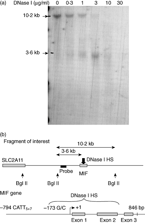
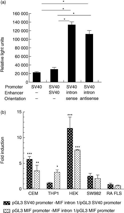
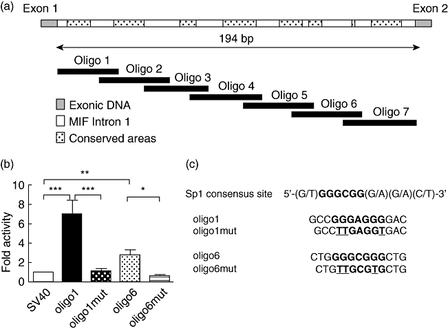
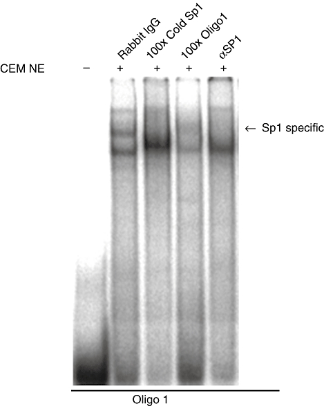
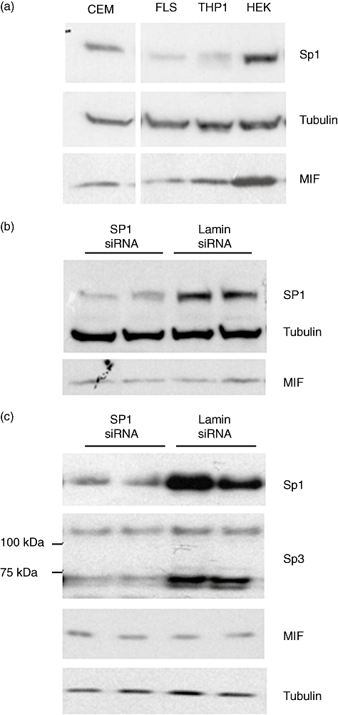
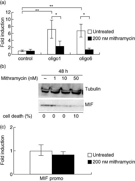
The aim of this study was to determine the genetic regulation of macrophage migration inhibitory factor (MIF). DNase I hypersensitivity was used to identify potential hypersensitive sites (HS) across the MIF gene locus. Reporter gene assays were performed in different human cell lines with constructs containing the native or mutated HS element. Following phylogenetic and transcription factor binding profiling, electrophoretic mobility shift assay (EMSA) and RNA interference were performed and the effects of incubation with mithramycin, an antibiotic that binds GC boxes, were also studied. An HS centred on the first intron of MIF was identified. The HS acted as an enhancer in human T lymphoblasts (CEMC7A), human embryonic kidney cells (HEK293T) and human monocytic cells (THP-1), but not in a fibroblast-like synoviocyte (FLS) cell line (SW982) or cultured FLS derived from rheumatoid arthritis (RA) patients. Two cis-elements within the first intron were found to be responsible for the enhancer activity. Mutation of the consensus Sp1 GC box on each cis-element abrogated enhancer activity and EMSA indicated Sp1 binding to one of the cis-elements contained in the intron. SiRNA knock-down of Sp1 alone or Sp1 and Sp3 together was incomplete and did not alter the enhancer activity. Mithramycin inhibited expression of MIF in CEMC7A cells. This effect was specific to the intronic enhancer and was not seen on the MIF promoter. These results identify a novel, cell type-specific enhancer of MIF. The enhancer appears to be driven by Sp1 or related Sp family members and is highly sensitive to inhibition via mithramycin.


Figures

Fig. 1
Fig. 1
Mapping of the DNase I hypersensitive site. (a) Genomic DNA was migrated overnight in a 0·8% agarose gel then transferred to a nylon membrane and hybridized overnight with a probe specific for the 10·2 Kb fragment of interest. Results are representative of two independent experiments. (b) Interpolation of hypersensitive site (HS) in untreated CEMC7A cells. The hypersensitive site spans ∼911 base pairs but is centred on the first intron.
Fig. 2
Fig. 2
The DNase I hypersensitive site is a cell type-specific enhancer. Reporter assays of the intronic constructs. (a) The first intron of the MIF gene was cloned into the pGL3 promoter vector (SV40) in both the sense and anti-sense orientation. As a positive control of enhancer activity the pGL3 control vector was used, which possesses an SV40 enhancer together with the SV40 promoter. The plasmids were transfected in CEMC7A cells and cells were left to incubate for 24 h before harvest and luciferase assay. *P< 0·0001. (b) The hypersensitive site is a cell type-specific enhancer. The transcriptional activity of the macrophage migration inhibitory factor (MIF) intron 1 enhancer was analysed in four different human cell lines: a T lymphoblast cell line (CEMC7A), an embryonic kidney cell line (HEK293T), a monocytic cell line (THP-1), a fibroblast-like synoviocyte cell line (FLS) and in primary fibroblast isolated from rheumatoid arthritis (RA) patients (RA FLS). Cells were transfected with constructs containing the MIF intron 1 sequence with either the SV40 promoter or the MIF promoter initiating luciferase gene expression. Following transfection, cells were incubated for 24 h before luciferase assays were performed. Results are representative of a minimum of two independent experiments (± standard error of the mean) performed in biological triplicates for all cell lines, and three different patients in biological duplicates for RA FLS. Results are expressed as a fold induction over the pGL3 SV40 or pGL3 MIF promoter vector. *P< 0·05; **P< 0·01; ***P< 0·001.
Fig. 3
Fig. 3
Mutation of Sp1 binding sites on cis-acting elements abrogates enhancer activity. (a) Schematic representation of the migration inhibitory factor (MIF) intronic enhancer and putative Sp1 binding sites. Seven oligonucleotides spanning the length of the intronic hypersensitive sites (HS) were cloned into pGL3 promoter. The hashed areas represent highly conserved Sp1 transcription factor binding sites. (b) Oligo-1 and oligo-6 are responsible for MIF enhancer activity. Cells were transfected with constructs containing wild-type (WT) or mutated oligo-1 or oligo-6 sequences. The pGL3 SV40 promoter-only construct was used as control and results are expressed as fold induction over the control. Following transfection, cells were incubated for 24 h before luciferase assays were performed. Results are representative of four to 10 independent experiments (±standard error of the mean) performed in single replicates. Results are expressed as a fold induction over the pGL3 SV40 or pGL3 MIF promoter vector. *P< 0·05; **P< 0·01; ***P< 0·001. (c) Sp1 binding site mutation. Three base pairs in each Sp1 consensus binding site were mutated from G to T, disrupting the core element for Sp1 binding.
Fig. 4
Fig. 4
Sp1 binds MIF intron 1. Electrophoretic mobility shift assay (EMSA) analysis demonstrating the binding of Sp1 to oligo-1. Nuclear extracts prepared from CEMC7A cells were incubated with radiolabelled oligo-1. Competition experiments were performed by the addition of 100× unlabelled oligonucleotide (cold Sp1) as indicated; 2 µg of rabbit IgG or Sp1 antibody added as indicated. Complexes were then resolved by native gel electrophoresis. Sp1 sequence specific complex marked by an arrow.
Fig. 5
Fig. 5
Neither Sp1 alone nor Sp1/Sp3 combined knock-down effects the enhancer activity. (a) Endogenous levels of Sp1 protein in different cell lines. Cell lysates of CEMC7A, FLS, THP-1 and HEK293T cells were immunoblotted for Sp1, alpha-tubulin and migration inhibitory factor (MIF). Results are representative of a minimum of two independent experiments performed in biological triplicates. (b) A pool of four siRNA sequences directed against Sp1 or Lamin A/C (negative control), 100 nM siRNA and 1–5 µg plasmid DNA were used per reaction. Transfected cells were incubated for 48 h Sp1 knock-down and endogenous MIF production was assessed by Western blotting. No down-regulation of the MIF protein was seen on endogenous MIF expression in HEK293T. (c) SiRNA directed against Sp1 (15 nM) and Sp3 (15 nM) or lamin A/C (negative control) (30 nM) were co-transfected into HEK293T cells. Cells were harvested 48 h post-transfection and subject to Western blot analysis with anti-Sp1, anti-Sp3, anti-MIF and anti-α-tubulin antibodies as indicated.
Fig. 6
Fig. 6
Mithramycin specifically inhibits the enhancer activity and endogenous migration inhibitory factor (MIF) expression. (a) Mithramycin inhibits the enhancer activity in CEMC7A cells. CEMC7A cells were transfected transiently either with a control vector possessing the SV40 promoter (control) or a vector containing oligo-1 or oligo-6. The cells were then treated with 200 nM mithramycin and left to incubate for 24 h. In untreated cells (white columns), the oligo-1 and oligo-6 reporter assays show enhancer activity. This activity was abrogated by the addition of mithramycin to the cells (black columns). Results are representative of a minimum of two independent experiments (±standard error of the mean) performed in biological triplicates. Results are expressed in fold induction over the untreated control vector. *P< 0·05; **P< 0·01. (b) Mithramycin inhibits endogenous expression of MIF in CEMC7A cells. CEMC7A cells were treated with increasing amounts of mithramycin (0, 1, 10 and 50 nM) for 24 h. Cell lysates were then collected and immunoblotted with the relevant primary anti-sera: anti-MIF goat antibody for MIF and anti-alpha-tubulin clone DM1A mouse antibody for tubulin. Cell death was counted by trypan blue exclusion. Results are representative of three independent experiments peformed in biological triplicates. (c) Mithramycin has no effect on the MIF promoter. CEMC7A cell were transfected with the MIF promoter only plasmid (MIF promo) and were left untreated (white column) or were treated with 200 nM mithramycin (black column) and left to incubate 24 h before luciferase assay was performed. Results are representative of two independent experiments performed in biological triplicates. Results are expressed in fold induction over untreated cells.

References

    1. Calandra T, Bernhagen J, Mitchell RA, Bucala R. The macrophage is an important and previously unrecognized source of macrophage migration inhibitory factor. J Exp Med. 1994;179:1895–902. - PMC - PubMed
    1. Bernhagen J, Calandra T, Mitchell RA, et al. MIF is a pituitary-derived cytokine that potentiates lethal endotoxaemia. Nature. 1993;365:756–9. - PubMed
    1. Bernhagen J, Bacher M, Calandra T, et al. An essential role for macrophage migration inhibitory factor in the tuberculin delayed-type hypersensitivity reaction. J Exp Med. 1996;183:277–82. - PMC - PubMed
    1. Kudrin A, Scott M, Martin S, et al. Human macrophage migration inhibitory factor: a proven immunomodulatory cytokine. J Biol Chem. 2006;281:29641–51. - PubMed
    1. Mikulowska A, Metz CN, Bucala R, Holmdahl R. Macrophage migration inhibitory factor is involved in the pathogenesis of collagen type II-induced arthritis in mice. J Immunol. 1997;158:5514–17. - PubMed

Publication types

MeSH terms