. 2010 Nov 18:10:253.
doi: 10.1186/1471-2229-10-253.

Classification of rice (Oryza sativa L. Japonica nipponbare) immunophilins (FKBPs, CYPs) and expression patterns under water stress

Affiliations

Classification of rice (Oryza sativa L. Japonica nipponbare) immunophilins (FKBPs, CYPs) and expression patterns under water stress

Jun Cheul Ahn et al. BMC Plant Biol. .

Abstract

Background: FK506 binding proteins (FKBPs) and cyclophilins (CYPs) are abundant and ubiquitous proteins belonging to the peptidyl-prolyl cis/trans isomerase (PPIase) superfamily, which regulate much of metabolism through a chaperone or an isomerization of proline residues during protein folding. They are collectively referred to as immunophilin (IMM), being present in almost all cellular organs. In particular, a number of IMMs relate to environmental stresses.

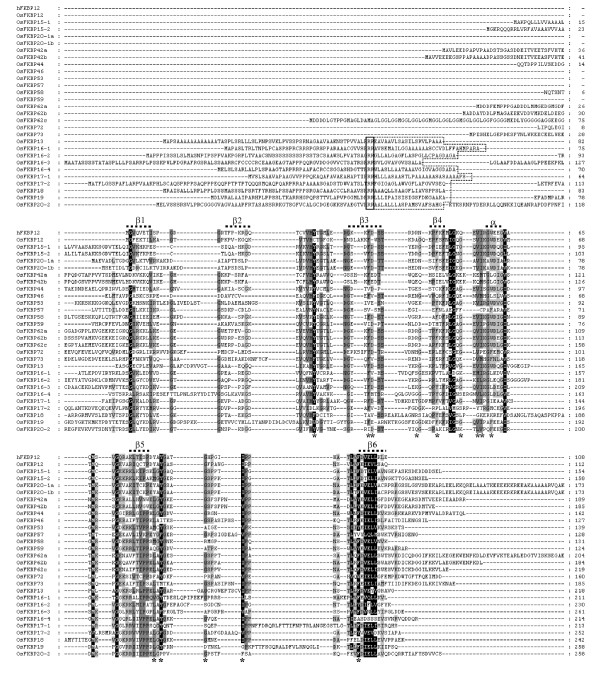
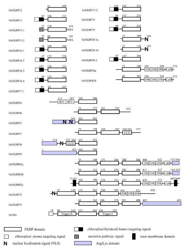
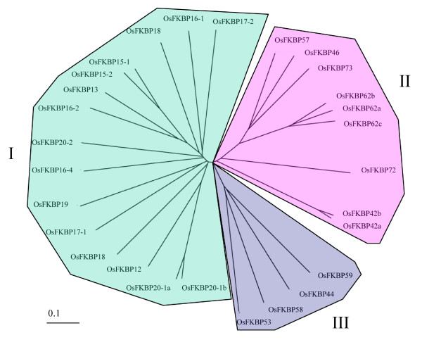
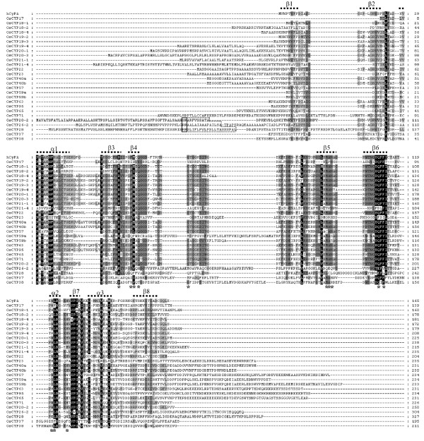
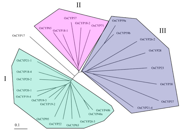
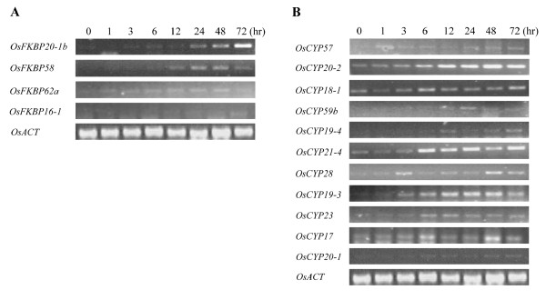
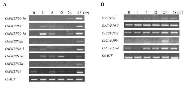
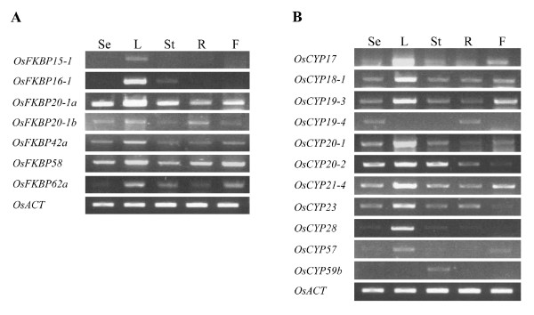
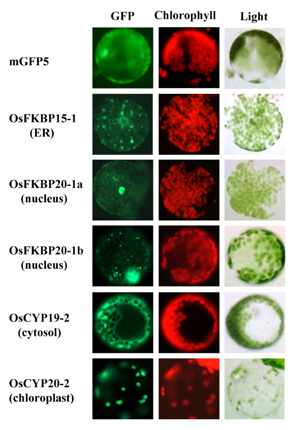
Results: FKBP and CYP proteins in rice (Oryza sativa cv. Japonica) were identified and classified, and given the appropriate name for each IMM, considering the ortholog-relation with Arabidopsis and Chlamydomonas or molecular weight of the proteins. 29 FKBP and 27 CYP genes can putatively be identified in rice; among them, a number of genes can be putatively classified as orthologs of Arabidopsis IMMs. However, some genes were novel, did not match with those of Arabidopsis and Chlamydomonas, and several genes were paralogs by genetic duplication. Among 56 IMMs in rice, a significant number are regulated by salt and/or desiccation stress. In addition, their expression levels responding to the water-stress have been analyzed in different tissues, and some subcellular IMMs located by means of tagging with GFP protein.

Conclusion: Like other green photosynthetic organisms such as Arabidopsis (23 FKBPs and 29 CYPs) and Chlamydomonas (23 FKBs and 26 CYNs), rice has the highest number of IMM genes among organisms reported so far, suggesting that the numbers relate closely to photosynthesis. Classification of the putative FKBPs and CYPs in rice provides the information about their evolutional/functional significance when comparisons are drawn with the relatively well studied genera, Arabidopsis and Chlamydomonas. In addition, many of the genes upregulated by water stress offer the possibility of manipulating the stress responses in rice.


Figures

Figure 1
Figure 1
Multiple sequence alignment of O. sativa FKBPs. Human FKBP12 (hFKBP12, GeneBank accession no. A35780) was used for determined for hFKBP12 were marked by asterisks (*). Secondary structure features (a-helix and b-sheets) derived from the hFKBP12 were displayed. The targeting signal of putative chloroplast (thylakoid lumen) localized OsFKBPs was manually adjusted (boxed): double Arg residues (--); the hydrophobic stretches following the double Args (---). Amino acids of full length protein for single FKBP domain OsFKBPs, and only the most conserved amino acids area for multiple FKBP domain OsFKBPs, were aligned. Backgrounds indicate percentage of amino acid similarity: black, 95%; dark grey, 70%; light grey, 40%.
Figure 2
Figure 2
Domain architecture of the O. sativa FKBP immunophilins. The beginning and ending amino acids numbers of each protein are shown at each end of the diagram. FKBP domains are represented by white box. The other functional domains such as tetrapeptide repeats (TPR), Arg/Lys amino acids rich domain (gray background box), coiled coil domain (CCD), internal repeats (RPT) and Calmodulin-binding motif (CaM) are indicated separately. The transmembrane domains (TM) that are not part of the signal peptide are labeled. The bipartite chloroplast/thylakoid targeting signal, ER Targeting signal, and the NLS are represented. The numbers above the boxes denote the amino acid positions of the function domains.
Figure 3
Figure 3
Unrooted phylogenetic trees of O. sativa FKBP proteins. The sequence alignment of Figure 1 (OsFKBPs) was used and the phylogenetic analysis was based on the sequence alignments by ClustalX http://align.genome.jp.
Figure 4
Figure 4
Multiple sequence alignment of O. sativa CYPs. Human CyPA (hCyPA, GeneBank accession no. P62937) was used for comparison. The amino acids necessary for CsA binding as determined for hCYPA were marked by asterisks (*). Secondary structure features (a-helix and b-sheets) derived from the hCYPA were displayed. The targeting signal of putative chloroplast (thylakoid lumen) localized OsCYPs was manually adjusted (boxed): double Arg residues (--); the hydrophobic stretches following the double Args (---). Amino acids of full length protein for single CYP domain OsCYPs, and only the most conserved amino acids area for multiple CYP domain OsCYPs, were aligned. Backgrounds indicate percentage of amino acid similarity: black, 95%; dark grey, 70%; light grey, 40%.
Figure 5
Figure 5
Domain architecture of the O. sativa CYP immunophilins. The beginning and ending amino acids numbers of each protein are shown at each end of the diagram. CYP domains are represented by white box. The other functional domains such as tetrapeptide repeats (TPR), Arg/Lys amino acids rich domain (gray background box), U-box, WD repeats (WD), RRM, Leu-zipper, and zinc-finger are indicated separately. The bipartite chloroplast/thylakoid targeting signal, ER Targeting signal, and the NLS are represented. The numbers above the boxes denote the amino acid positions of the function domains.
Figure 6
Figure 6
Unrooted phylogenetic trees of O. sativa CYP proteins. The sequence alignment of Figure 4 (OsCYPs) was used and the phylogenetic analysis was based on the sequence alignments by ClustalX http://align.genome.jp.
Figure 7
Figure 7
Expression level of representative immunophilins under salt stress. Semi-quantitative RT-PCR was carried out with gene specific primers using cDNAs from RNA samples. RNAs were isolated from two-week-old seedlings were exposed to 200 mM NaCl and tissues were harvested at the indicated time points. The rice actin level was used for control. A, OsFKBPs RT-PCR analysis; B, OsCYPs RT-PCR analysis.
Figure 8
Figure 8
Expression level of representative immunophilins under desiccation stress. Semi-quantitative RT-PCR was performed with gene specific primers using cDNAs from RNA samples. RNAs were isolated from two-week-old seedlings were exposed to drought stress and tissues were harvested at the indicated time points. The rice actin level was used for control. A, OsFKBPs RT-PCR analysis; B, OsCYPs RT-PCR analysis.
Figure 9
Figure 9
Expression levels of some immunophilins (especially, showing changes in mRNA expression levels under water stress) in different tissues. Semi-quantitative RT-PCR was performed with gene specific primers using cDNAs from RNA samples. RNAs were isolated from RNA samples isolated from different tissues. The tissues were used 6 days growing seedling (Se), leaves (L), stems (St), roots (R) and flowers (F). The rice actin level was used for control. A, OsFKBPs RT-PCR analysis; B, OsCYPs RT-PCR analysis.
Figure 10
Figure 10
Subcellular localization of O. sativa immunophilins using GFP fusion proteins. The full length ORF of Os immunophilins FKBP15-2, FKBP20-1a, FKBP20-1b, CYP19-2 and CYP20-2 were fused to the N-terminal mGFP5 in pCAMBIA1302 and expressed in N. benthamiana leaf cell transformed using Agrobacterium. Localization of fluorescent signals was examined at 48 h after Agrobacterium infiltration under fluorescence microscopy. The images of GFP, chlorophyll auto-fluorescence and bright-field images are shown.

Similar articles

Cited by

References

    1. Schreiber SL. Chemistry and biology of the immuophilins and their immuno-suppressive ligands. Science. 1991;251:283–287. doi: 10.1126/science.1702904. - DOI - PubMed
    1. Fisher G, Wittmann-Liebold B, Lang K, Kiefhaber T, Schmid FX. Cyclophilin and the peptidyl-prolyl cis-trans isomerase are probably identical proteins. Nature. 1989;337:476–478. doi: 10.1038/337476a0. - DOI - PubMed
    1. Reimer U, Fischer G. Local structural changes caused by peptidyl-prolyl cis/trans isomerization in the native state of proteins. Biophys Chem. 2002;96:203–212. doi: 10.1016/S0301-4622(02)00013-3. - DOI - PubMed
    1. He Z, Li L, Luan S. Immunophilins and parvulins. Superfamily of peptidyl prolyl isomerases in Arabidopsis. Plant Physiol. 2004;134:1248–1267. doi: 10.1104/pp.103.031005. - DOI - PMC - PubMed
    1. Pemberton TJ, Kay JE. Identification and comparative analysis of the peptidyl-prolyl cis/trans isomerase repetoires of H. sapiens, D. melanogaster, C. elegans, S. cerevisiae &Sz. Pombe. Comp Funct Genomics. 2005;6:277–300. doi: 10.1002/cfg.482. - DOI - PMC - PubMed

Publication types

MeSH terms