. 2010 Nov 18:7:81.
doi: 10.1186/1742-2094-7-81.

Hippocampal FGF-2 and BDNF overexpression attenuates epileptogenesis-associated neuroinflammation and reduces spontaneous recurrent seizures

Affiliations

Hippocampal FGF-2 and BDNF overexpression attenuates epileptogenesis-associated neuroinflammation and reduces spontaneous recurrent seizures

Roberta Bovolenta et al. J Neuroinflammation. .

Abstract

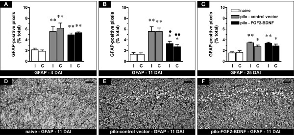
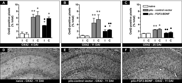
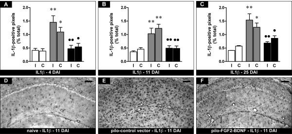
Under certain experimental conditions, neurotrophic factors may reduce epileptogenesis. We have previously reported that local, intrahippocampal supplementation of fibroblast growth factor-2 (FGF-2) and brain-derived neurotrophic factor (BDNF) increases neurogenesis, reduces neuronal loss, and reduces the occurrence of spontaneous seizures in a model of damage-associated epilepsy. Here, we asked if these possibly anti-epileptogenic effects might involve anti-inflammatory mechanisms. Thus, we used a Herpes-based vector to supplement FGF-2 and BDNF in rat hippocampus after pilocarpine-induced status epilepticus that established an epileptogenic lesion. This model causes intense neuroinflammation, especially in the phase that precedes the occurrence of spontaneous seizures. The supplementation of FGF-2 and BDNF attenuated various parameters of inflammation, including astrocytosis, microcytosis and IL-1β expression. The effect appeared to be most prominent on IL-1β, whose expression was almost completely prevented. Further studies will be needed to elucidate the molecular mechanism(s) for these effects, and for that on IL-1β in particular. Nonetheless, the concept that neurotrophic factors affect neuroinflammation in vivo may be highly relevant for the understanding of the epileptogenic process.


Figures

Figure 1
Figure 1
Effects of FGF-2 and BDNF on hippocampal GFAP-positive cells (putative astrocytes). (A-C) Time course of alterations in GFAP expression, expressed as percent of GFAP-positive pixels in naïve rats (open bars), pilocarpine rats inoculated with the control vector (pilo - control vector, gray bars) or with the vector expressing FGF-2 and BDNF (pilo - FGF2-BDNF, solid bars) in the right, ipsilateral to inoculation (I) and in the left, contralateral (C) hippocampus. Analysis has been performed 4 (A), 11 (B) and 25 days (C) after inoculation (DAI), i.e. 7, 14 and 28 days after pilocarpine SE. Data are presented as mean ± s.e.m. for 6 animals per group. *P < 0.05, **P < 0.01 versus naïve; P < 0.05, ●●P < 0.01 versus pilo-control vector. One-way ANOVA and post-hoc Newman-Keuls test. (D-F) Representative sections showing GFAP immunohistochemistry in the CA1 region of naïve rats (D), of pilocarpine rats killed 11 days after control vector injection (E) and of pilocarpine rats 11 days after FGF2-BDNF vector injection (F). The pattern of changes observed in CA1 was identical for the entire hippocampus. Horizontal bar = 50 μm.
Figure 2
Figure 2
Effects of FGF-2 and BDNF on hippocampal Ox42-positive cells (putative microglia). (A-C) Time course of the alterations in Ox42 expression, expressed as percent of Ox42-positive pixels. See Figure 1 for details. Data are presented as mean ± s.e.m. for 6 animals per group. *P < 0.05, **P > 0.01 versus naïve; P < 0.05, ●●P < 0.01 versus pilo-control vector. One-way ANOVA and post-hoc Newman-Keuls test. (D-F) Representative sections showing Ox42 immunohistochemistry in the CA1 region of naïve rats (D), of pilocarpine rats killed 11 days after control vector injection (E) and of pilocarpine rats 11 days after FGF2-BDNF vector injection (F). The pattern of changes observed in CA1 was identical for the entire hippocampus. Horizontal bar = 50 μm.
Figure 3
Figure 3
Effects of FGF-2 and BDNF on hippocampal IL1β-positive cells. (A-C) Time course of the alterations in IL1β expression, expressed as percent of IL1β-positive pixels. See Figure 1 for details. Data are presented as mean ± s.e.m. for 6 animals per group. *P < 0.05, **P < 0.01 versus naïve; P < 0.05, ●●P > 0.01 versus pilo-control vector. One-way ANOVA and post-hoc Newman-Keuls test. (D-F) Representative sections showing IL1β immunohistochemistry in the CA1 region of naïve rats (D), of pilocarpine rats killed 11 days after control vector injection (E) and of pilocarpine rats 11 days after FGF2-BDNF vector injection (F). The pattern of changes observed in CA1 was identical for the entire hippocampus. Horizontal bar = 50 μm.

References

    1. Pitkanen A, Sutula TP. Is epilepsy a progressive disorder? Prospects for new therapeutic approaches in temporal-lobe epilepsy. Lancet Neurol. 2002;1:173–181. doi: 10.1016/S1474-4422(02)00073-X. - DOI - PubMed
    1. Temkin NR. Antiepileptogenesis and seizure prevention trials with antiepileptic drugs: meta-analysis of controlled trials. Epilepsia. 2001;42:515–524. doi: 10.1046/j.1528-1157.2001.28900.x. - DOI - PubMed
    1. Simonato M, Tongiorgi E, Kokaia M. Angels and demons: neurotrophic factors and epilepsy. Trends Pharmacol Sci. 2006;27:631–638. doi: 10.1016/j.tips.2006.10.002. - DOI - PubMed
    1. Schinder AF, Poo M. The neurotrophin hypothesis for synaptic plasticity. Trends Neurosci. 2000;23:639–645. doi: 10.1016/S0166-2236(00)01672-6. - DOI - PubMed
    1. Paradiso B, Marconi P, Zucchini S, Berto E, Binaschi A, Bozac A, Buzzi A, Mazzuferi M, Magri E, Navarro Mora G, Rodi D, Su T, Volpi I, Zanetti L, Marzola A, Manservigi R, Fabene PF, Simonato M. Localized delivery of fibroblast growth factor-2 and brain-derived neurotrophic factor reduces spontaneous seizures in an epilepsy model. Proc Natl Acad Sci USA. 2009;106:7191–7196. doi: 10.1073/pnas.0810710106. - DOI - PMC - PubMed

Publication types

Substances