. 2010 Nov 18:9:295.
doi: 10.1186/1476-4598-9-295.

Effects of human mesenchymal stem cells on ER-positive human breast carcinoma cells mediated through ER-SDF-1/CXCR4 crosstalk

Affiliations

Effects of human mesenchymal stem cells on ER-positive human breast carcinoma cells mediated through ER-SDF-1/CXCR4 crosstalk

Lyndsay V Rhodes et al. Mol Cancer. .

Abstract

Background: Adult human mesenchymal stem cells (hMSC) have been shown to home to sites of carcinoma and affect biological processes, including tumour growth and metastasis. Previous findings have been conflicting and a clear understanding of the effects of hMSCs on cancer remains to be established. Therefore, we set out to investigate the impact of hMSCs on the oestrogen receptor positive, hormone-dependent breast carcinoma cell line MCF-7.

Results: In this study, we show the effects of hMSCs on cancer cells are mediated through a secreted factor(s) which are enhanced by cancer cell-hMSC contact/communication. In addition to enhanced proliferation when in co-culture with hMSCs, MCF-7 cells were found to have increased migration potential in vitro. Inhibition of ER signalling by the pure anti-oestrogen ICI 182,780 decreased the effect of hMSCs on MCF-7 cell proliferation and migration supporting a role for ER signalling in the hMSC/MCF-7 cell interaction. Additionally, hMSCs have been shown to secrete a wide variety of growth factors and chemokines including stromal cell-derived factor-1 (SDF-1). This coupled with the knowledge that SDF-1 is an ER-mediated gene linked with hormone-independence and metastasis led to the investigation of the SDF-1/CXCR4 signalling axis in hMSC-MCF-7 cell interaction. Experiments revealed an increase in SDF-1 gene expression both in vivo and in vitro when MCF-7 cells were cultured with hMSCs. SDF-1 treatment of MCF-7 cells alone increased proliferation to just below that seen with hMSC co-culture. Additionally, blocking SDF-1 signalling using a CXCR4-specific inhibitor decreased hMSC induced proliferation and migration of MCF-7. However, the combined treatment of ICI and AMD3100 reduced MCF-7 cell proliferation and migration below control levels, indicating targeting both the ER and CXCR4 pathways is effective in decreasing the hMSCs induction of MCF-7 cell proliferation and migration.

Conclusions: The sum of these data reveals the relationship between tumour microenvironment and tumour growth and progression. Better understanding of the mechanisms involved in this tumour stroma cell interaction may provide novel targets for the development of treatment strategies for oestrogen receptor positive, hormone-independent, and endocrine-resistant breast carcinoma.


Figures

Figure 1
Figure 1
hMSCs enhance MCF-7 cell proliferation. (A) MCF-7-GFP cells cultured with or without hMSCs over 72 hours. Cells were fixed and stained with anti-Ki-67 (red) and nuclei were counter stained with DAPI (blue). Representative images of MCF-7 cells cultured alone (left) or with hMSCs (right) at 400×. (B) Quantification of GFP cells positive for Ki-67 staining from 10 fields of view per treatment. Data is represented as percent positive MCF-7 cells as compared to total number of MCF-7 cells normalized to MCF-7 control group. (C) MCF-7 cells were seeded in the lower chamber of a 24-well plate and MCF-7 cells (control) or hMSCs were seeded on the upper transwell insert. After 7 days of culture, inserts were removed and MTT was added to each well. After 4 hours, cells were solubilized and absorbance read. (D) MCF-7 cells were treated with MCF-7 (control) or hMSC conditioned media for 24 hours. After 4 hours of MTT treatment, cells were solubilized and absorbance read. All data are represented as percent proliferation normalized to control treated cells ± SEM, (* p < 0.05, *** p < 0.001).
Figure 2
Figure 2
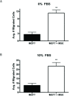
hMSCs increase migration of MCF-7 cells in vitro. MCF-7-GFP cells alone or in combination with hMSCs (1:1) were seeded in the upper chamber of a transwell system and treated with DMSO. Lower wells contained either (A) serum-free culture media (0%) or (B) culture media supplemented with 10% FBS (10%). After 48 hours cells were fixed and the number of GFP-positive migrated cells counted. Bars represent average number of migrated cell per condition ± SEM, (** p < 0.01).
Figure 3
Figure 3
Inhibition of ER signaling decreases hMSC stimulated MCF-7 proliferation and migration in vitro. (A) MCF-7 cells were seeded in the lower chamber of a 24-well plate with MCF-7 cells or hMSCs were seeded in the upper well. Upper and lower chambers were treated with either DMSO (control) or ICI (100 nM). After 7 days of culture, inserts were removed and MTT was added to each well. Cells were solubilized and absorbance read. All data are represented as normalized percent proliferation compared to MCF-7 + DMSO (control) treated cells ± SEM, (* p < 0.05, ** p < 0.01). (B) MCF-7-GFP cells alone or in combination with hMSCs (1:1) were seeded in the upper chamber of a transwell system and lower wells contained culture media supplemented with 10% FBS (10%). Upper and lower chambers were treated with either DMSO (control) or ICI (100 nM). After 48 hours cells were fixed and the number of GFP positive migrated cells counted. Bars represent average number of migrated cell per condition ± SEM, (* p < 0.05, *** p < 0.001).
Figure 4
Figure 4
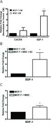
hMSCs enhance SDF-1 gene transcription in vivo and in vitro. (A) MCF-7 cells were treated with conditioned media from MCF-7 cells (control) or hMSCs for 24 hours. Cells were then harvested for PCR analysis of CXCR4 and SDF-1. (B) Endpoint oestrogen and matrigel tumours were harvested at day 36 post cell injection and gene expression analyzed by qPCR. (C) Endpoint matrigel tumours grown in the absence of oestrogen were harvested at day 44 and gene expression analyzed by qPCR. Data represent fold change relative to control as normalized to internal β-actin ± SEM, (*, p < 0.05).
Figure 5
Figure 5
Inhibition of CXCR4 signaling decreases hMSC stimulated MCF-7 proliferation and migration in vitro. (A) MCF-7 cells cultured alone were treated with DMSO (control) or SDF-1 (50 ng/ml) for 72 hours. Cells were fixed and stained with anti-Ki-67 (red) and nuclei were counter stained with DAPI (blue). Representative images of MCF-7 cells at 200×. (B) Quantification of cells positive for Ki-67 staining from 10 fields of view per treatment. Data is represented as percent positive cells of total cells counted. Bars represent mean values ± SEM. (C) MCF-7 cells were seeded in the lower chamber of a 24-well plate and a transwell insert was placed in each well. MCF-7 cells or hMSCs were seeded in the upper well. Upper and lower chambers were treated with DMSO (control) or AMD3100 (5 μg/ml). After 7 days of culture, inserts were removed and MTT was added to each well. After 4 hours cells were lysed and absorbance read. All data are represented as mean percent proliferation as compared to MCF-7 + DMSO (control) treated cells ± SEM. (D) MCF-7-GFP cells alone or in combination with hMSCs (1:1) were seeded in the upper chamber of a transwell system where the lower wells contained culture media supplemented with 10% FBS (10%). Upper and lower chambers were treated with DMSO (control) or AMD3100 (5 μg/ml). After 48 hours cells were fixed and the number of GFP-positive migrated cells counted. Bars represent average number of migrated cell per condition ± SEM, (*, p < 0.05; **, p < 0.01).
Figure 6
Figure 6
Co-inhibition of ER and CXCR4 signaling decreases hMSC stimulated MCF-7 proliferation and migration in vitro. MCF-7-GFP cells cultured with or without hMSCs were treated with DMSO (control) or ICI (100 nM) + AMD3100 (5 μg/ml) for 72 hours. Cells were fixed and stained with anti-Ki-67 (red) and nuclei were counter stained with DAPI (blue). (A) Representative images of MCF-7 cells cultured alone (left) or with hMSCs (right) at 200×. (B) Quantification of GFP cells positive for Ki-67 staining from 10 fields of view per treatment. Data is represented as percent positive MCF-7 cells as compared to total number of MCF-7 cells counted. Bars represent mean values ± SEM. (C) MCF-7 cells alone or in combination with hMSCs (1:1 mix) were seeded in the upper chamber of a transwell system where the lower wells contained culture media supplemented with 10% FBS (10%). Upper and lower chambers were treated with either DMSO (control) or ICI (100 nM) + AMD3100 (5 μg/ml). After 48 hours cells were fixed and the number of migrated cells counted. Bars represent average number of migrated cell per condition ± SEM, (*, p < 0.05; ***, p < 0.001).

References

    1. Ali S, Coombes RC. Estrogen receptor alpha in human breast cancer: occurrence and significance. J Mammary Gland Biol Neoplasia. 2000;5:271–281. doi: 10.1023/A:1009594727358. - DOI - PubMed
    1. Jordan VC, Gottardis MM, Robinson SP, Friedl A. Immune-deficient animals to study "hormone-dependent" breast and endometrial cancer. J Steroid Biochem. 1989;34:169–176. doi: 10.1016/0022-4731(89)90079-4. - DOI - PubMed
    1. Brodie A, Sabnis G, Jelovac D. Aromatase and breast cancer. J Steroid Biochem Mol Biol. 2006;102:97–102. doi: 10.1016/j.jsbmb.2006.09.002. - DOI - PMC - PubMed
    1. Jordan VC. Long-term tamoxifen therapy to control or to prevent breast cancer: laboratory concept to clinical trials. Prog Clin Biol Res. 1988;262:105–123. - PubMed
    1. Reed MJ. The role of aromatase in breast tumors. Breast Cancer Res Treat. 1994;30:7–17. doi: 10.1007/BF00682737. - DOI - PubMed

Publication types

MeSH terms