. 2010 Nov 19:3:109.
doi: 10.1186/1756-3305-3-109.

The nature and combination of subunits used in epitope-based Schistosoma japonicum vaccine formulations affect their efficacy

Affiliations

The nature and combination of subunits used in epitope-based Schistosoma japonicum vaccine formulations affect their efficacy

Xuefeng Wang et al. Parasit Vectors. .

Abstract

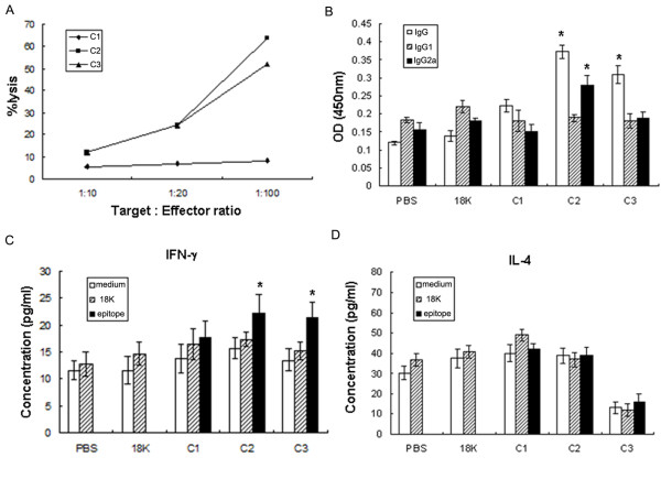
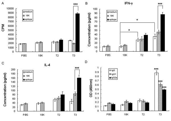
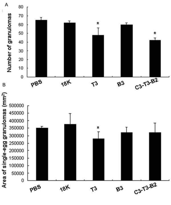
Background: Schistosomiasis remains a major public health problem in endemic countries and is caused by infections with any one of three primary schistosome species. Although there are no vaccines available to date, this strategy appears feasible since natural immunity develops in individuals suffering from repeated infection during a lifetime. Since vaccinations resulting in both Th1- and Th2-type responses have been shown to contribute to protective immunity, a vaccine formulation with the capacity for stimulating multiple arms of the immune response will likely be the most effective. Previously we developed partially protective, single Th- and B cell-epitope-based peptide-DNA dual vaccines (PDDV) (T3-PDDV and B3-PDDV, respectively) capable of eliciting immune responses against the Schistosoma japonicum 22.6 kDa tegument antigen (Sj22.6) and a 62 kDa fragment of myosin (Sj62), respectively.

Results: In this study, we developed PDDV cocktails containing multiple epitopes of S. japonicum from Sj22.6, Sj62 and Sj97 antigens by predicting cytotoxic, helper, and B-cell epitopes, and evaluated vaccine potential in vivo. Results showed that mice immunized with a single-epitope PDDV elicited either Tc, Th, or B cell responses, respectively, and mice immunized with either the T3- or B3- single-epitope PDDV formulation were partially protected against infection. However, mice immunized with a multicomponent (3 PDDV components) formulation elicited variable immune responses that were less immunoprotective than single-epitope PDDV formulations.

Conclusions: Our data show that combining these different antigens did not result in a more effective vaccine formulation when compared to each component administered individually, and further suggest that immune interference resulting from immunizations with antigenically distinct vaccine targets may be an important consideration in the development of multicomponent vaccine preparations.


Figures

Figure 1
Figure 1
CTL-PDDVs induced the cytotoxicity effect and produced antibody. (A) Effect of C-PDDV vaccination on cytotoxicity. Seven days after the last C-PDDV, 18K-PDDV, or PBS immunization epitope-specific cytotoxic activity was measured by incubating murine spleen effector cells or p815 target cells with either C1-, C2-, or C3-18K fusion peptides, 18K control peptide, or medium only, then mixed cells at E:T rations ranging from 1:10 to 1:100. The CTL activity of the cells was tested using Na2[51Cr]O4 assay. Data are expressed as the mean ± SD (n = 6 per group) of 18 mice from three independent experiments performed in triplicate wells. (B) Serum antibody subtype profile following C-PDDV vaccination. Whole IgG, IgG1, and IgG2a responses to SWA (0.1 mg/ml) following vaccination with C-PDDV formulations, or controls were measured by ELISA. Data are expressed as the mean ± SD (n = 6 per group) of 18 mice from three independent experiments performed in triplicate wells. * P < 0.05 and ** P < 0.01, compared to the 18K-PDDV and PBS groups. (C-D) Cytokine profile analysis following C-PDDV vaccination. IFN-γ (C) and IL-4 (D) production of splenocytes harvested from every vaccination group were determined by culturing in triplicate. In 96-well plates, 106cells/well were cultured for 48 h in 200 μl of media in the presence of C1-, C2-, C3-18K fusion peptide (10 μg/ml), 18K (10 μg/ml), or media alone. Supernatants were collected after 48 h of culture for cytokine detection. Bars show the mean ± SD (n = 6 per group) of 18 mice from three independent experiments performed in triplicate wells.
Figure 2
Figure 2
T3-PDDV induced both cellular and humoral immune responses. (A) T-PDDV induced cellular immunity. Seven days after the last immunization with T-PDDVs, 18K-PDDV, or PBS, splenocytes were harvested and antigen-specific proliferation was measured. Splenocytes (2 × 10⁵/well) from each mouse were incubated in triplicate for three days in 200 μl in 96-well plates in the presence of either the T2-18K, T3-18K, or 18K peptides (10 μg/ml), or media alone. To each well 0.5 μCi [3H] thymidine was added 16 h before the end of the incubation period. Data are expressed as the mean ± SD (n = 6 per group) of 18 mice from three independent experiments performed in triplicate wells. *** P < 0.001. (B-C) Cytokine production following T2- or T3-PDDV vaccinations. IFN-γ (B) and IL-4 (C) production was measured in splenocytes harvested and cultured from the respective vaccination groups for 48 h. Bars show the mean ± SD (n = 6 per group) of 18 mice from three independent experiments performed in triplicate wells. * P < 0.05; *** P < 0.001. (D) IgG, IgG1, and IgG2a responses in immunized mice. Antibody responses to SWA (0.1 mg/ml) were determined by ELISA. *** P < 0.001, compared with 18K-PDDV and PBS groups. Data are expressed as the mean ± SD (n = 6 per group) of 18 mice from three independent experiments performed in triplicate wells.
Figure 3
Figure 3
B3-PDDV induced the highest antibody response. (A) Analysis of B-PDDV-induced antibody responses. Seven days after the last immunization with B-PDDVs, 18K-PDDV, or PBS, mouse whole IgG, IgG1, and IgG2a antibody responses to SWA (0.1 mg/ml) were analyzed by ELISA. Data are expressed as the mean ± SD (n = 6 per group) of 18 mice from three independent experiments performed in triplicate wells. ** P < 0.01 and *** P < 0.001, compared to the 18K-PDDV and PBS groups. (B) Splenocyte proliferation assay. Splenocytes (2 × 10⁵/well) from each mouse were incubated in triplicate wells for three days in 200 μl of media in 96-well plates in the presence of B1-, B2-, B3-18K fusion peptide, or 18K (10 μg/ml) peptides (or media alone). Proliferation was determined by measuring [3H] thymidine incorporation for the last 16 h of the experiment. Data are expressed as the mean ± SD (n = 6 per group) of 18 mice from three independent experiments. * P < 0.05. (C-D) Cytokine production analysis. Supernatants were collected after 48 h of culture and examined for IFN-γ (C) or IL-4 (D). Bars show the mean ± SD (n = 6 per group) of 18 mice from three independent experiments performed in triplicate wells.
Figure 4
Figure 4
Cytokines and antibodies responses in mice vaccinated with CTL-, T-, and B-PDDV cocktails. (A-B) Cytokine responses following vaccination with C-, T- and B-PDDV multicomponent formulations. Seven days after the last immunization with either multicomponent PDDV formulations, 18K-PDDV, or PBS, splenocyte production of IFN-γ (A) and IL-4 (B) from each vaccine group was assessed. Splenocytes from each mouse were cultured (106/well) in triplicate for 48 h in the presence or absence of SWA (50 μg/ml). Supernatants were collected after 48 h and IFN-γ and IL-4 production was assessed. The data are expressed as the mean ± SD (n = 6 per group) of 12 mice from two independent experiments performed in triplicate wells. (C-E) Antibody responses following multicomponent vaccinations. IgG (C), IgG1 (D), and IgG2a (E) responses to SWA in immunized mice were determined by ELISA. Data are expressed as the mean ± SD (n = 6 per group) of 12 mice from two independent experiments performed in triplicate wells. *P < 0.05 and ***P < 0.001, compared to the 18K-PDDV and PBS groups.
Figure 5
Figure 5
Histopathology of egg granulomas in the livers. (A) Six weeks after challenge, mice were sacrificed. After portal perfusion, livers were dissected and stained with H&E for microscopic examination. The number of granulomas in the liver of each mouse was counted in 10 random fields and the data are expressed as the mean ± SD of 16 mice (8 mice/group) from two independent experiments. (B) The size of nonconfluent granulomas formed around a single egg was assessed using a video micrometer. The data are expressed as the mean ± SD of 16 mice (8 mice/group) from two independent experiments. *P < 0.05, compared to the 18K-PDDV and PBS groups.

Similar articles

Cited by

References

    1. Hotez PJ, Brindley PJ, Bethony JM, King CH, Pearce EJ, Jacobson J. Helminth infections: the great neglected tropical diseases. J Clin Invest. 2008;118:1311–1321. doi: 10.1172/JCI34261. - DOI - PMC - PubMed
    1. King CH. Toward the elimination of schistosomiasis. N Engl J Med. 2009;360:106–109. doi: 10.1056/NEJMp0808041. - DOI - PubMed
    1. Doenhoff MJ, Cioli D, Utzinger J. Praziquantel: mechanisms of action, resistance and new derivatives for schistosomiasis. Curr Opin Infect Dis. 2008;21:659–667. doi: 10.1097/QCO.0b013e328318978f. - DOI - PubMed
    1. Fenwick A, Webster JP. Schistosomiasis: challenges for control, treatment and drug resistance. Curr Opin Infect Dis. 2006;19:577–582. doi: 10.1097/01.qco.0000247591.13671.6a. - DOI - PubMed
    1. Balen J, Zhao ZY, Williams GM, McManus DP, Raso G, Utzinger J, Zhou J, Li YS. Prevalence, intensity and associated morbidity of Schistosoma japonicum infection in the Dongting Lake region, China. Bull World Health Organ. 2007;85:519–526. doi: 10.2471/BLT.06.034033. - DOI - PMC - PubMed

LinkOut - more resources