Review
. 2011 May;125(1-2):95-104.
doi: 10.1016/j.jsbmb.2010.11.004. Epub 2010 Nov 16.

Inhibitors of type 5 17β-hydroxysteroid dehydrogenase (AKR1C3): overview and structural insights

Affiliations
Review

Inhibitors of type 5 17β-hydroxysteroid dehydrogenase (AKR1C3): overview and structural insights

Michael C Byrns et al. J Steroid Biochem Mol Biol. 2011 May.

Abstract

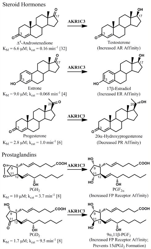
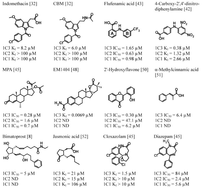
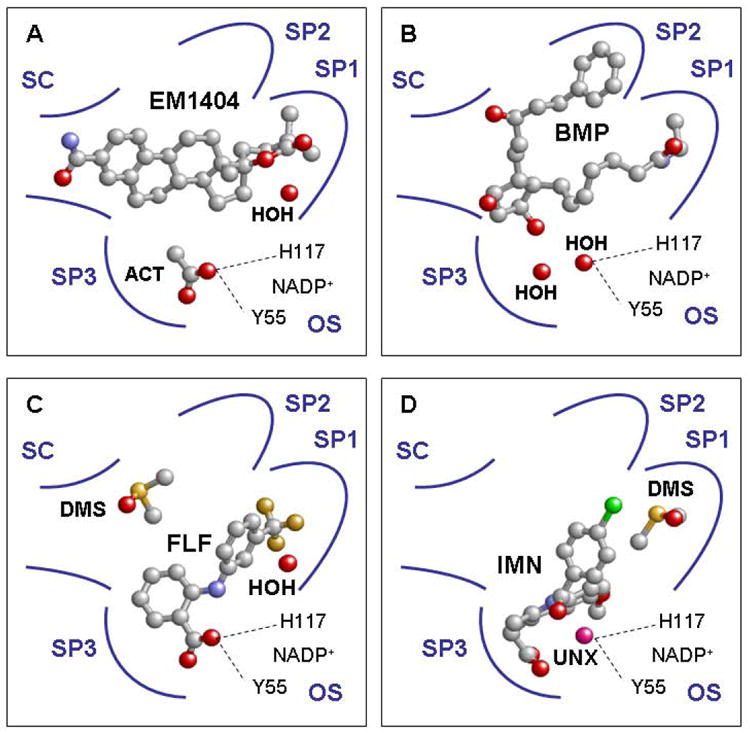
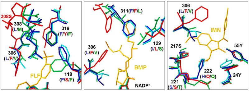
There is considerable interest in the development of an inhibitor of aldo-keto reductase (AKR) 1C3 (type 5 17β-hydroxysteroid dehydrogenase and prostaglandin F synthase) as a potential therapeutic for both hormone-dependent and hormone-independent cancers. AKR1C3 catalyzes the reduction of 4-androstene-3,17-dione to testosterone and estrone to 17β-estradiol in target tissues, which will promote the proliferation of hormone dependent prostate and breast cancers, respectively. AKR1C3 also catalyzes the reduction of prostaglandin (PG) H(2) to PGF(2α) and PGD(2) to 9α,11β-PGF(2), which will limit the formation of anti-proliferative prostaglandins, including 15-deoxy-Δ(12,14)-PGJ(2), and contribute to proliferative signaling. AKR1C3 is overexpressed in a wide variety of cancers, including breast and prostate cancer. An inhibitor of AKR1C3 should not inhibit the closely related isoforms AKR1C1 and AKR1C2, as they are involved in other key steroid hormone biotransformations in target tissues. Several structural leads have been explored as inhibitors of AKR1C3, including non-steroidal anti-inflammatory drugs, steroid hormone analogues, flavonoids, cyclopentanes, and benzodiazepines. Inspection of the available crystal structures of AKR1C3 with multiple ligands bound, along with the crystal structures of the other AKR1C isoforms, provides a structural basis for the rational design of isoform specific inhibitors of AKR1C3. We find that there are subpockets involved in ligand binding that are considerably different in AKR1C3 relative to the closely related AKR1C1 or AKR1C2 isoforms. These pockets can be used to further improve the binding affinity and selectivity of the currently available AKR1C3 inhibitors. Article from the special issue on Targeted Inhibitors.


Figures

Figure 1
Figure 1
Reactions catalyzed by AKR1C3. AKR1C3 will enhance proliferative signaling in various hormone-responsive cells through the formation of androgens and estrogens with increased affinity for the androgen receptor (AR) and estrogen receptor (ER), respectively, as well as through reduction of progesterone to a metabolite with decreased affinity for the progesterone receptor (PR). AKR1C3 will also catalyze prostaglandin (PG) reduction reactions that enhance proliferative pathways by activating the F prostanoid (FP) receptor and preventing formation of anti-proliferative 15-deoxy-Δ12,14-PGJ2, which forms through spontaneous dehydration and rearrangement of PGD2.
Figure 2
Figure 2
Chemical structures of representative AKR1C3 inhibitors and their inhibitory potency towards AKR1C3 and closely related isoforms AKR1C1 and AKR1C2.ND, not determined; CBM, 4-chlorobenzoyl melatonin; MPA, medroxyprogesterone acetate. The A- and B-rings of flufenamic acid are indicated.
Figure 3
Figure 3
Occupancies of AKR1C3 ligand binding sub-sites by inhibitors EM1404, bimatoprost, flufenamic acid, and indomethacin (A through D). SC =steroid channel; OS = oxyanion site; ACT = acetate ion; HOH = water; BMP = bimatoprost; FLF = flufenamic acid; DMS = dimethyl sulfoxide; IMN = indomethacin; UNX = unknown atom or ion; SP1- 3 = subpockets 1-3. A, the lactone portion of EM1404 is bound in SP1 while the estrone portion extends into the steroid channel (PDB ID:1ZQ5[48]). B, the α-chain of bimatoprost is bound in SP1 while the β-chain of the inhibitor is bound in SP2 (PDB ID:2F38[58]). C, the trifluoromethyl substituted B ring of flufenamic acid is located in SP1 and the carboxylic acid moiety of the moleculeisanchored at the oxyanion site (PDB ID:1S2C[56]). D, the bridge carbonyl group of indomethacin is positioned via an unknown atom located at the oxyanion site. The p-chlorobenzoyl group of indomethacin projects into SP1, while the indole ring and carboxylic acid is bound in the well defined SP3 (PDB ID:1S2A[56]). Ligands are depicted in ball-and-stick representation and the following atomic coloring scheme: C = grey; O = red; N = blue; F = orange; S = yellow; Cl = green; unknown = pink.
Figure 4
Figure 4
Comparison of SP1, SP2, and SP3 sub-pockets of four human AKR1C enzymes. The crystal structures used for comparisons are AKR1C1•NADP+•20α-hydroxyprogesterone (PDB ID:1MRQ)[62], AKR1C2•NADP+•ursodeoxycholate (PDB ID:1IHI)[61], and AKR1C4•NADP+ (PDB ID:2FVL). The respective AKR1C3 structures in the left, middle and right panels are AKR1C3•NADP+•flufenamic acid (PDB ID:1S2C, for comparison of SP1), AKR1C3•NADP+•bimatoprost (PDB ID:2F38, for comparison of SP2) and AKR1C3•NADP+•indomethacin (PDB ID:1S2A, for comparison of SP3). The residues are shown in stick presentation and in cyan for AKR1C1, blue for AKR1C2, red for AKR1C3, and green for AKR1C4. For clarity, only key residues for binding are shown and are labeled by the number and their respective identities in AKR1C2 (blue), AKR1C3 (red), and AKR1C4 (green). AKR1C1 has the same residues as AKR1C2 at all positions shown. Ligands are shown in gold. FLF = flufenamic acid, BMP = bimatoprost, and IMN = indomethacin.Left, AKR1C3 has a larger SP1 site than the other isoforms due to different mainchain position at 308 and different residues at 118. In addition, S118 of AKR1C3 can form sidechain hydrogen bonding interaction with a ligand, whereas the corresponding residue F118 in other isoforms cannot. Middle, structural differences at positions 129 and 311 make the SP2 pocket shorter for AKR1C1, AKR1C2 and AKR1C4 than for AKR1C3. S129 of AKR1C3 can form sidechain hydrogen bonding interactions with a ligand, whereas the corresponding I129 of AKR1C1/2 and L129 of AKR1C4 cannot. Right, the sidechain of F306 in AKR1C3 assumes a conformation that exposes the SP3 site for indomethacin binding. However, the corresponding L306 of AKR1C1/2 and V306 for AKR1C4 with more rigid sidechains would extend into the indole ring of indomethacin.

References

    1. Penning TM. Human hydroxysteroid dehydrogenases and pre-receptor regulation: Insights into inhibitor design and evaluation. J Steroid Biochem Mol Biol. 2010 current issue. - PMC - PubMed
    1. Marchais-Oberwinkler S, Hennl C, Möller G, Klein T, Negri M, Oster A, Spadaro A, Werth R, Wetzel M, Xu K, Frotscher M, Hartmann RW, Adamski J. 17β-Hydroxysteroid dehydrogenase (17β-HSD): gene, protein structures, novel therapeutic targets and recent progress in inhibitor development. J Steroid Biochem Mol Biol. 2010 current issue. - PubMed
    1. Poirier D. Contribution to the development of inhibitors of 17β-hydroxysteroid dehydrogenase types 1 and 7: key tools for studying and treating estrogen-dependent diseases. J Steroid Biochem Mol Biol. 2010 current issue. - PubMed
    1. Byrns MC, Duan L, Lee SH, Blair IA, Penning TM. Aldo-keto reductase 1C3 expression in MCF-7 cells reveals roles in steroid hormone and prostaglandin metabolism that may explain its over-expression in breast cancer. J Steroid Biochem Mol Biol. 2010;118:177–187. - PMC - PubMed
    1. Dufort I, Rheault P, Huang X-F, Soucy P, Luu-The V. Characteristics of a highly labile human type 5 17β-hydroxysteroid dehydrogenase. Endocrinology. 1999;140:568–574. - PubMed

Publication types

MeSH terms