

Degeneration of the mid-cingulate cortex in amyotrophic lateral sclerosis detected in vivo with MR spectroscopy
- PMID: 21087934
- PMCID: PMC7965735
- DOI: 10.3174/ajnr.A2289
Degeneration of the mid-cingulate cortex in amyotrophic lateral sclerosis detected in vivo with MR spectroscopy
Abstract
Background and purpose: Various lines of evidence implicate cerebral involvement beyond the motor cortex in ALS, including the cingulate gyrus and the thalamus. The purpose of this study was to assess neurodegeneration in these regions in vivo by using MRSI.
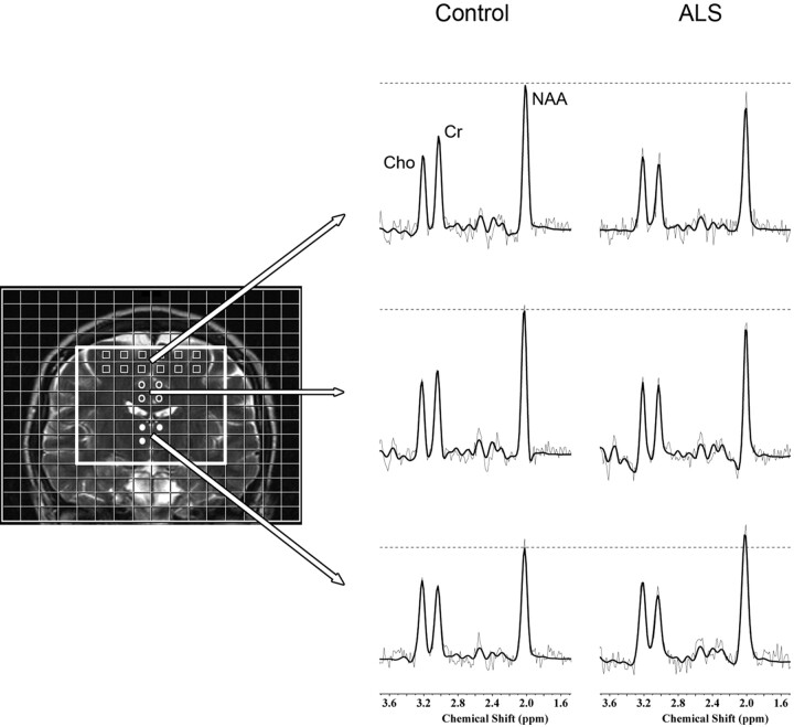
Materials and methods: Fourteen patients with ALS and 14 healthy controls underwent MRSI by using a coronal acquisition scheme. The NAA/Cho ratio was quantified in the MCC, thalamus, and motor cortex (PCG).
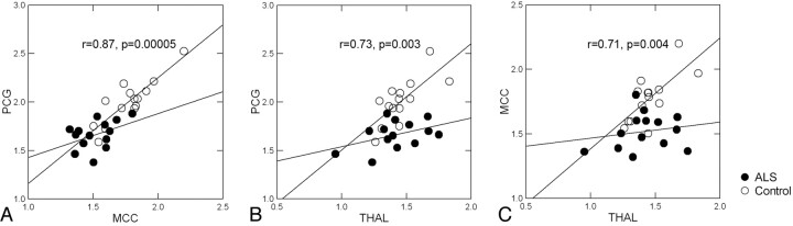
Results: NAA/Cho was reduced in the MCC in patients with ALS compared with the controls (P = .0004). There was no difference in NAA/Cho in the thalamus (P = .59). We also found a strong correlation of NAA/Cho among the PCG, MCC, and the thalamus in controls, which was absent in patients with ALS.
Conclusions: Neurodegeneration beyond the motor cortex is present in the MCC in ALS. The significant correlation of NAA/Cho among the PCG, MCC, and the thalamus in healthy subjects likely reflects the neuronal connectivity among these regions. The loss of these relationships in patients with ALS suggests that such connectivity is not responsible for the pattern of degeneration in these regions.
Figures
References
-
- Ringholz GM, Appel SH, Bradshaw M, et al. . Prevalence and pattern of cognitive impairment in sporadic ALS. Neurology 2005;65:586–90 - PubMed
-
- Strong MJ. The syndromes of frontotemporal dysfunction in amyotrophic lateral sclerosis. Amyotroph Lateral Scler 2008;9:323–38 - PubMed
-
- Kiernan JA, Hudson AJ. Frontal lobe atrophy in motor neuron diseases. Brain 1994;117(pt 4):747–57 - PubMed
-
- Ikemoto A, Hirano A, Akiguchi I. Neuropathology of amyotrophic lateral sclerosis with extra-motor system degeneration: characteristics and differences in the molecular pathology between ALS with dementia and Guamanian ALS. Amyotroph Lateral Scler Other Motor Neuron Disord 2000;1:97–104 - PubMed
-
- Ellis CM, Suckling J, Amaro E, Jr, et al. . Volumetric analysis reveals corticospinal tract degeneration and extramotor involvement in ALS. Neurology 2001;57:1571–78 - PubMed
Publication types
MeSH terms
Substances
LinkOut - more resources
Full Text Sources
Medical
Miscellaneous