

Optimization and initial experience of a multisection balanced steady-state free precession cine sequence for the assessment of fetal behavior in utero
- PMID: 21087938
- PMCID: PMC7965695
- DOI: 10.3174/ajnr.A2295
Optimization and initial experience of a multisection balanced steady-state free precession cine sequence for the assessment of fetal behavior in utero
Abstract
Background and purpose: The assessment of motor function is an essential component of neurologic examinations, which imaging studies have extended to the fetus. US assessment is hampered by a limited FOV, whereas MR imaging has the potential to be an alternative. Our objectives were to optimize a cine MR imaging sequence for capturing fetal movements and to perform a pilot analysis of the relationship between the frequency of movements and uterine spatial constrictions in healthy fetuses.
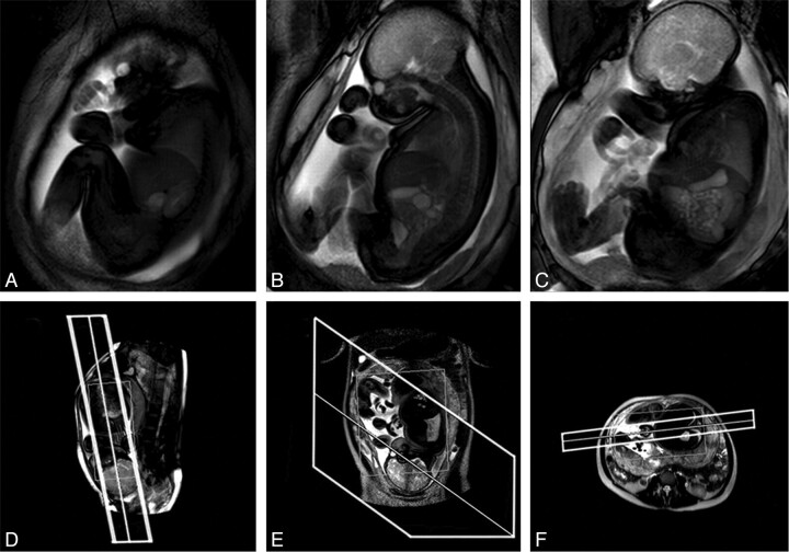
Materials and methods: Initially, a bSSFP cine sequence was selected for optimization, and various compromises were explored in all acquisition parameters to achieve an effective balance between anatomic coverage of the fetus and the temporal resolution of cine data, with the aim of maximizing both. Subsequently, cross-sectional qualitative and quantitative analyses of fetal movements were performed prospectively by using a cohort of 37 healthy fetuses (median GA, 29 weeks; range, 20-37 weeks) with the optimized cine protocol. Two smaller subgroups were selected for representative sampling of overall behavior patterns by using cine data of longer duration and for volumetric quantification of free intrauterine space.
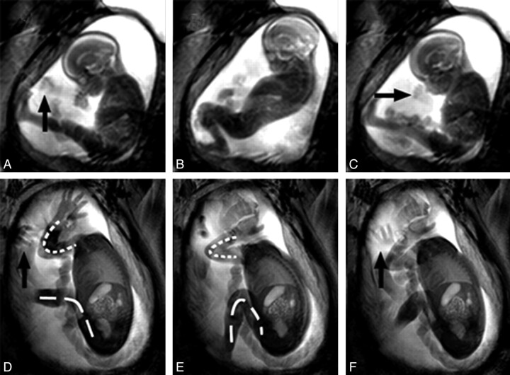
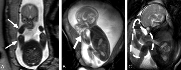
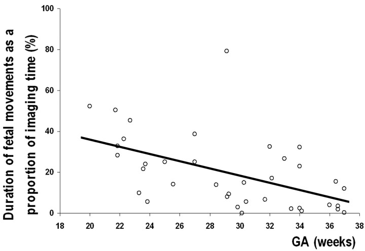
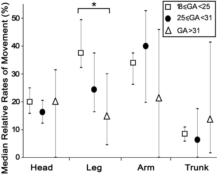
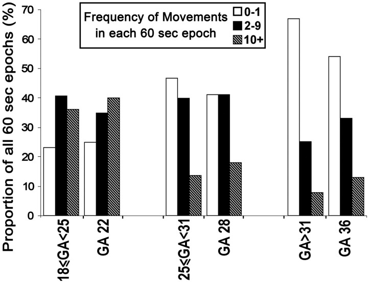
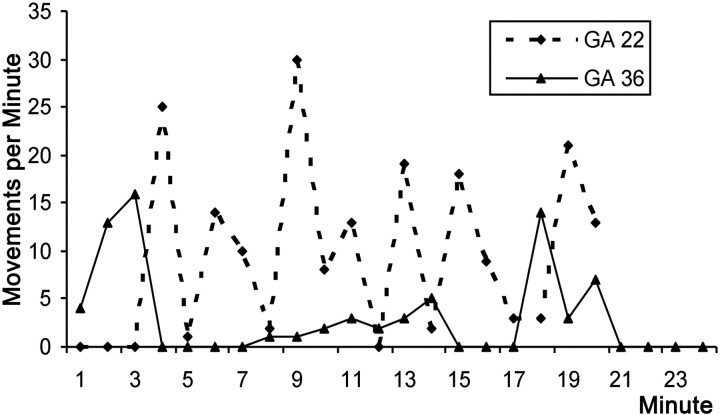
Results: The optimized cine sequence, with TR/TE of 3.21/1.59 ms, coupled with parallel imaging and partial-Fourier imaging, resulted in a section-acquisition time of 0.303 seconds. Anatomic coverage was enhanced by using a combination of thick sagittal sections (30-40 mm) and multisection acquisitions to display movements in all fetal limbs, head, and trunk simultaneously. All expected motor patterns were observed throughout this gestational period, and a significant decreasing trend in overall movement frequency with age was demonstrated (r = -0.514, P = .0011). Also a significant negative correlation was found between overall movement frequency and the total intrauterine free space (r = -0.703, P = .0001). Furthermore, a significant decrease in the frequency of leg movements was shown in fetuses older then 30 weeks' GA compared with those younger than that (P = .015).
Conclusions: Cine MR imaging is effective for observing fetal movements from midgestation with near full-body coverage. Also, reductions in free space with increasing GA appear to be a factor in the gradual reductions in overall levels of fetal activity as well as in restrictions in movement within specific regions of the fetal anatomy.
Figures

References
-
- Einspieler C, Prechtl HF. Prechtl's assessment of general movements: a diagnostic tool for the functional assessment of the young nervous system. Ment Retard Dev Disabil Res Rev 2005;11:61–67 - PubMed
-
- Spittle AJ, Doyle LW, Boyd RN. A systematic review of the clinimetric properties of neuromotor assessments for preterm infants during the first year of life. Dev Med Child Neurol 2008;50:254–66. Epub 2008 Jan 7 - PubMed
-
- Murney ME, Campbell SK. The ecological relevance of the Test of Infant Motor Performance elicited scale items. Phys Ther 1998;78:479–89 - PubMed
-
- Cioni G, Ferrari F, Einspieler C, et al. Comparison between observation of spontaneous movements and neurologic examination in preterm infants. J Pediatr 1997;130:704–11 - PubMed
-
- de Vries JI, Visser GH, Prechtl HF. The emergence of fetal behaviour. II. Quantitative aspects. Early Hum Dev 1985;12:99–120 - PubMed
Publication types
MeSH terms
Grants and funding
LinkOut - more resources
Full Text Sources
Medical