. 2011 Feb;32(2):413-8.
doi: 10.3174/ajnr.A2290. Epub 2010 Nov 18.

Acute hyperammonemic encephalopathy in adults: imaging findings

Affiliations

Acute hyperammonemic encephalopathy in adults: imaging findings

J M U-King-Im et al. AJNR Am J Neuroradiol. 2011 Feb.

Abstract

Background and purpose: Acute hyperammonemic encephalopathy has significant morbidity and mortality unless promptly treated. We describe the MR imaging findings of acute hyperammonemic encephalopathy, which are not well-recognized in adult patients.

Materials and methods: We retrospectively reviewed the clinical and imaging data and outcome of consecutive patients with documented hyperammonemic encephalopathy seen at our institution. All patients underwent cranial MR imaging at 1.5T.

Results: Four patients (2 women; mean age, 42 ± 13 years; range, 24-55 years) were included. Causes included acute fulminant hepatic failure, and sepsis with a background of chronic hepatic failure and post-heart-lung transplantation with various systemic complications. Plasma ammonia levels ranged from 55 to 168 μmol/L. Bilateral symmetric signal-intensity abnormalities, often with associated restricted diffusion involving the insular cortex and cingulate gyrus, were seen in all cases, with additional cortical involvement commonly seen elsewhere but much more variable and asymmetric. Involvement of the subcortical white matter was seen in 1 patient only. Another patient showed involvement of the basal ganglia, thalami, and midbrain. Two patients died (1 with fulminant cerebral edema), and 2 patients survived (1 neurologically intact and the other with significant intellectual impairment).

Conclusions: The striking common imaging finding was symmetric involvement of the cingulate gyrus and insular cortex in all patients, with more variable and asymmetric additional cortical involvement. These specific imaging features should alert the radiologist to the possibility of acute hyperammonemic encephalopathy.


Figures

Fig 1.
Fig 1.
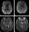
Patient 1. A 24-year-old woman with complicated (cardiac failure, pulmonary edema, chest infection, sternal wound sepsis, and venous thrombosis) and very protracted postoperative course in the ICU post–heart-lung transplantation. She was on a standard immunosuppressive regimen and had been on total parenteral nutrition. Five weeks after surgery, she deteriorated neurologically with seizures and decreased level of consciousness. Her plasma ammonium level was 168 μmol/L. DWI (A and B) and FLAIR images (C and D) show symmetric abnormal signal intensity in the insular (arrows) and cingulate cortices (arrowheads) bilaterally. Extensive signal-intensity change involving both the cortex and subcortical white matter is also seen in right temporo-occipital and left occipital lobes.
Fig 2.
Fig 2.
Patient 1. Follow-up brain CT scan after 7 days in patient 1, who continued to deteriorate neurologically despite therapeutic measures. Repeat plasma ammonium level decreased to 110 μmol/L. A, Effacement of suprasellar cisterns in keeping with bilateral uncal herniation is seen. B, Image at a level above the lateral ventricles shows widespread loss of cortical sulci and gray-white differentiation, in keeping with cerebral edema. The patient died soon after the study was performed.
Fig 3.
Fig 3.
Patient 2. A 48-year-old man with fulminant acute hepatic failure due to acetaminophen overdose with a background of chronic alcohol abuse, His ICU stay was complicated by sepsis due to pneumonia and Clostridium difficile diarrhea. On day 8 post−ICU admission, he deteriorated neurologically with a decreased level of consciousness, abnormal posturing, and seizures. DWI (A and B) and T2-weighted images (C and D) show symmetric abnormal high signal intensity in the insular and cingulate cortices (open arrows). High signal intensity is also seen involving the cortex of the temporoparietal lobes bilaterally, bilateral thalami (arrowheads), and brain stem (closed arrow). His plasma ammonium level was 102 μmol/L. The patient's condition deteriorated further, and he died the next day without repeat imaging.
Fig 4.
Fig 4.
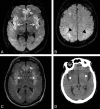
Patient 3. A 55-year-old man with severe sepsis of unknown origin (positive blood cultures for Enterococcus species) with a background of schizophrenia, chronic hepatitis C, portal hypertension, and esophageal varices. The patient had a reduced level of consciousness and seizures. His plasma ammonium level was 82 μmol/L. A−C, DWI (A and B) and FLAIR image (C) show high signal intensity in the insular and cingulate cortices (arrows) and the perirolandic cortex bilaterally (black arrowheads). There is also subtle high signal intensity in the subinsular regions. With conservative therapeutic measures, the patient gradually improved neurologically, his plasma ammonium level decreased to 50 μmol/L, but he had significant residual intellectual impairment. D, Follow-up brain CT after 1 month showed widening of Sylvian fissures bilaterally (white arrowheads) in keeping with insular atrophy.
Fig 5.
Fig 5.
Patient 4. A 42-year-old woman was admitted with sepsis related to her central dialysis line, with a background of liver transplant, chronic graft rejection, and hepatorenal syndrome. Her immunosuppressant regimen included tacrolimus and cyclosporin. Her condition deteriorated with respiratory failure, hypotension, seizures, and a drop in the GCS score. Her plasma ammonium level was 55 μmol/L. FLAIR image (A) and DWI (B) show abnormal signal intensity in the cortex involving the bilateral insular and cingulate and left posterior temporal lobes (arrows). The patient made an excellent recovery without significant neurologic deficit with supportive therapeutic measures.

Comment in

References

    1. Clay AS, Hainline BE. Hyperammonemia in the ICU. Chest 2007;132:1368–78 - PubMed
    1. Lichtenstein GR, Yang YX, Nunes FA, et al. . Fatal hyperammonemia after orthotopic lung transplantation. Ann Intern Med 2000;132:283–87 - PubMed
    1. Blaser S, Feigenbaum A. A neuroimaging approach to inborn errors of metabolism. Neuroimaging Clin N Am 2004;14:307–29, ix - PubMed
    1. Bindu PS, Sinha S, Taly AB, et al. . Cranial MRI in acute hyperammonemic encephalopathy. Pediatr Neurol 2009;41:139–42 - PubMed
    1. Sato S, Yokota C, Toyoda K, et al. . Hyperammonemic encephalopathy caused by urinary tract infection with urinary retention. Eur J Intern Med 2008;19:e78–79 - PubMed

MeSH terms

LinkOut - more resources