. 2011 Mar 1;52(3):1573-85.
doi: 10.1167/iovs.10-6413.

Polarized secretion of PEDF from human embryonic stem cell-derived RPE promotes retinal progenitor cell survival

Affiliations

Polarized secretion of PEDF from human embryonic stem cell-derived RPE promotes retinal progenitor cell survival

Danhong Zhu et al. Invest Ophthalmol Vis Sci. .

Abstract

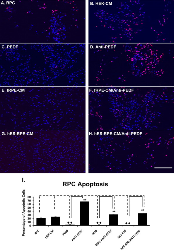
Purpose: Human embryonic stem cell-derived RPE (hES-RPE) transplantation is a promising therapy for atrophic age-related macular degeneration (AMD); however, future therapeutic approaches may consider co-transplantation of hES-RPE with retinal progenitor cells (RPCs) as a replacement source for lost photoreceptors. The purpose of this study was to determine the effect of polarization of hES-RPE monolayers on their ability to promote survival of RPCs.

Methods: The hES-3 cell line was used for derivation of RPE. Polarization of hES-RPE was achieved by prolonged growth on permeable inserts. RPCs were isolated from 16- to 18-week-gestation human fetal eyes. ELISA was performed to measure pigment epithelium-derived factor (PEDF) levels from conditioned media.

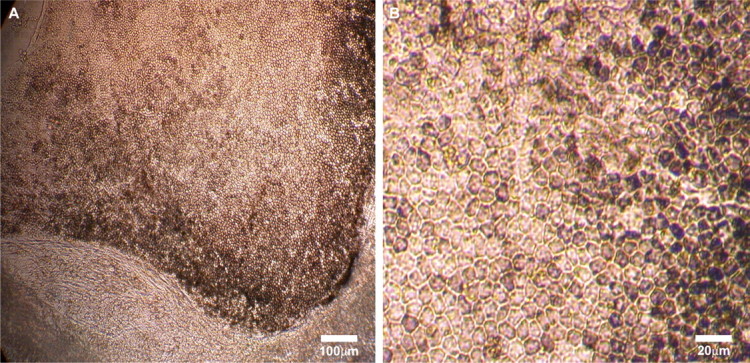
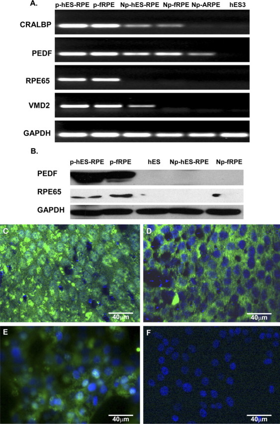
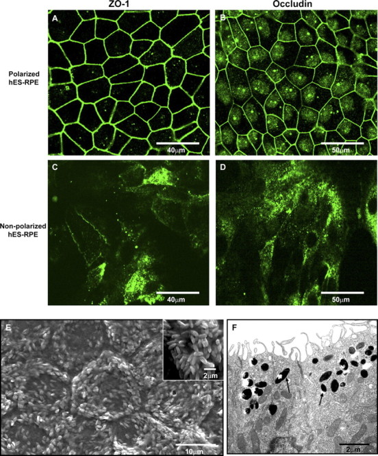
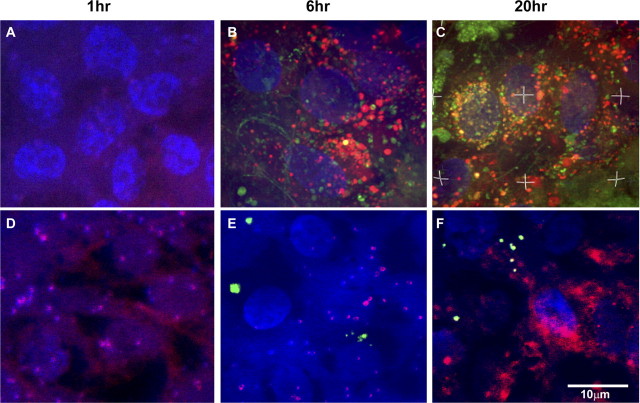
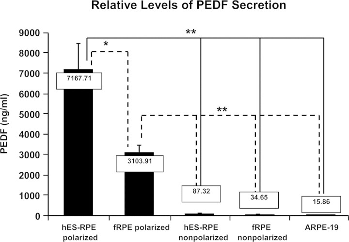
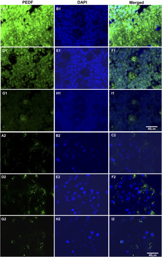
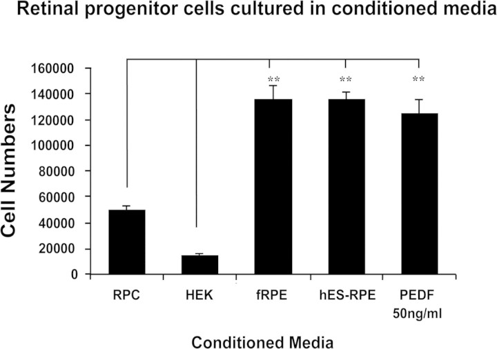
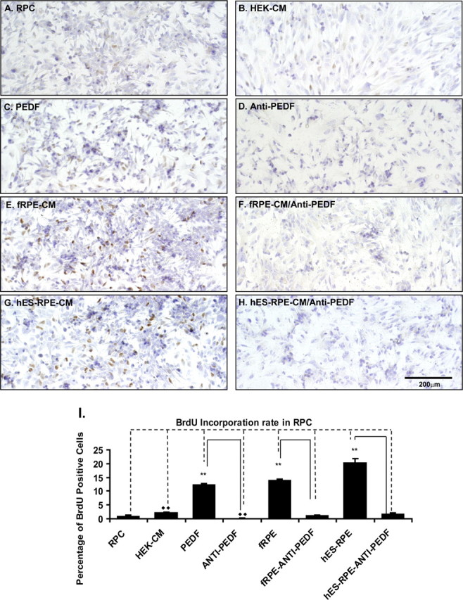
Results: Pigmented RPE-like cells appeared as early as 4 weeks in culture and were subcultured at 8 weeks. Differentiated hES-RPE had a normal chromosomal karyotype. Phenotypically polarized hES-RPE cells showed expression of RPE-specific genes. Polarized hES-RPE showed prominent expression of PEDF in apical cytoplasm and a marked increase in secretion of PEDF into the medium compared with nonpolarized culture. RPCs grown in the presence of supernatants from polarized hES-RPE showed enhanced survival, which was ablated by the presence of anti-PEDF antibody.

Conclusions: hES-3 cells can be differentiated into functionally polarized hES-RPE cells that exhibit characteristics similar to those of native RPE. On polarization, hES-RPE cells secrete high levels of PEDF that can support RPC survival. These experiments suggest that polarization of hES-RPE would be an important feature for promotion of RPC survival in future cell therapy for atrophic AMD.


Figures

Figure 1.
Figure 1.
Phase-contrast microscopic images of hES-RPE cells derived from hES-3 stem cell line. After neural precursor induction and RPE spontaneous differentiation, the pigmented hexagonal RPE cells appeared as early as 4 weeks in our culture conditions and reached a high enough cell count for subculture at approximately 8 weeks. The hES-RPE cells could be subcultured for 5 to 6 passages and still retain their pigmented hexagonal morphology. (A) RPE-like cells differentiated from one embryoid body and cultured in the differentiation medium for 8 weeks. (B) Passage 3 hES-RPE cells cultured for 4 weeks.
Figure 2.
Figure 2.
RT-PCR, Western blot analysis, and immunofluorescent staining of RPE-specific genes expressed by hES-RPE derived from hES-3 embryonic stem cell line and control cells. Polarized hES-RPE (p-hES-RPE; passage 3) and polarized fetal human RPE (p-fRPE) were compared with nonpolarized hES-RPE (Np-hES-RPE; passage 3), nonpolarized fetal human RPE (Np-fRPE), nonpolarized ARPE-19 (Np-ARPE), and undifferentiated hES3 cells. The polarized hES-RPE expressed high levels of RPE-specific gene products. (A) RT-PCR for CRALBP, PEDF, RPE65, and VMD2; GAPDH was the loading control. (B) Western blot for PEDF and RPE65 with GAPDH as loading control. (CF) Immunofluorescent staining. PEDF was highly expressed in polarized hES-RPE cells at passage 3 (C) and less prominently expressed in nonpolarized hES-RPE cells (E). Immunofluorescent staining for RPE65 was found in polarized RPE cells (D), but not in nonpolarized hES-RPE cells (F). Immunoreactivity was detected using secondary antibodies conjugated to FITC (green). Nuclei are counterstained with DAPI (blue).
Figure 3.
Figure 3.
Confocal immunofluorescent, scanning electron microscopic (SEM), and transmission electron microscopic (TEM) images of polarized hES-RPE (passage 3) derived from the hES-3 cell line. The highly polarized hES-RPE cells exhibited positive immunofluorescent staining of the tight junction specific proteins ZO-1 and occludin (A, B). Although focal expression of ZO-1 and occludin was detectable in nonpolarized hES-RPE cells, the proteins did not localize to the cell membrane (C, D). Immunoreactivity was detected using secondary antibodies conjugated to FITC (green). Nuclei are counterstained with DAPI (blue). The polarized sheet of hES-RPE showed apical localization of microvilli on SEM (E; inset: one cell at higher magnification). TEM image of two cells in a polarized hES-RPE sheet shows apical microvilli, melanin granules in apical cytoplasm (black arrows) and apical tight junctions (white arrow) between the two cells (F).
Figure 4.
Figure 4.
Confocal images to show the phagocytosis of ROS by polarized hES-RPE (passage 3) derived from hES-3 stem cell line compared to nonpolarized hES-RPE. The polarized and nonpolarized hES-RPE cells were challenged with FITC-labeled ROS (green) at different time points. Lysosomes were labeled with fluorescent dye (red), and nuclei were labeled with DAPI (blue). The phagocytosis assay revealed that there were no FITC-ROS containing phagosomes in either polarized or nonpolarized hES-RPE cells after a 1-hour challenge (A, D). FITC-ROS containing phagosomes appeared in polarized hES-RPE cells as early as 6 hours after ROS challenge (B), whereas they did not appear in nonpolarized hES-RPE at the same time period after ROS challenge (E). After 20 hours of FITC-ROS treatment, the labeled phagosomes reached the highest numbers inside polarized hES-RPE cells and many of them fused with red lysosomes (yellow; C), whereas few FITC-ROS containing phagosomes were found inside nonpolarized hES-RPE cells (F). Images are representative of experiments performed in triplicate.
Figure 5.
Figure 5.
ELISA analysis of PEDF secretion by hES-RPE (passage 3) derived from hES-3 embryonic stem cell line and control cells. All PEDF values in the supernatants were normalized to total protein levels of the corresponding cellular lysates. Numerical values of PEDF concentration are shown in the box at the top of each bar. The polarized hES-RPE and the polarized fRPE secreted approximately 100-fold greater levels of PEDF than the nonpolarized hES-RPE, fRPE, and ARPE19 cells. ELISA revealed that polarized hES-RPE secreted twice as much PEDF as that of polarized fRPE. Results represent three independent experiments. *P < 0.05; **P < 0.01.
Figure 6.
Figure 6.
Confocal images of PEDF immunofluorescence in polarized (A1–I1) and nonpolarized (A2–I2) hES-RPE (passage 3) taken from equivalent levels in the apical region (AC), middle region (DF), and basal region (GI). The majority of PEDF is localized to the apical cytoplasm of polarized hES-RPE cells (A1–I1), whereas PEDF was reduced in amount and found more in the central region in the cytoplasm of nonpolarized hES-RPE cells (A2–I2). Immunoreactivity for PEDF was detected using secondary antibodies conjugated to FITC (green). Nuclei are counterstained with DAPI (blue). Merged images show both PEDF immunoreactivity and DAPI nuclear staining. Images are representative of three independent experiments.
Figure 7.
Figure 7.
RPC counting. Human RPCs cultured in the polarized hES-RPE (passage 3) CM, polarized fRPE CM, or RPC medium with 50 ng/mL PEDF supplement had a significantly higher number of cells than those cultured in the control HEK-CM or RPC medium. Results represent three independent experiments; **P < 0.01.
Figure 8.
Figure 8.
BrdU incorporation analysis of RPC proliferation after culture in various CM. The RPCs cultured in the polarized hES-RPE (passage 3) CM, polarized fRPE CM, or RPC medium with 50 ng/mL PEDF supplement had more proliferating cells (BrdU-positive cells) than those cultured in the regular RPC medium, HEK-CM, polarized fRPE CM, or hES-RPE CM, with anti-PEDF antibody. (AH) Light microscopy images of BrdU-positive RPCs. (A) RPCs cultured in the regular RPC medium; (B) RPCs in HEK-CM; (C) RPCs in RPC medium with 50 ng/mL PEDF; (D) RPCs in RPC medium with 50 ng/mL PEDF and 20 μg/mL anti-PEDF antibody; (E) RPCs in the polarized fRPE CM; (F) RPCs in the polarized fRPE CM with 20 μg /mL anti-PEDF antibody; (G) RPCs in the polarized hES-RPE CM; (H) RPCs in the polarized hES-RPE CM with 20 μg/mL anti-PEDF antibody. (I) The quantification of BrdU-positive RPCs from three individual experiments. The RPCs cultured in the polarized hES-RPE CM, polarized fRPE CM, or RPC medium with recombinant PEDF supplement had a significantly higher number of BrdU-positive cells than those cultured in the regular RPC medium, HEK-CM, polarized fRPE- CM or hES-RPE CM, and recombinant PEDF with anti-PEDF antibody. **P < 0.01.
Figure 9.
Figure 9.
Apoptosis (TUNEL) assay to measure RPC death in various CM after overnight challenge with 250 μM H2O2. The RPCs cultured in the polarized hES-RPE (passage 3) CM, polarized fRPE CM, or RPC medium with 50 ng/mL PEDF supplement showed less cell death than those cultured in the regular RPC medium, HEK-CM, polarized fRPE CM, or hES-RPE CM with anti-PEDF antibody. Images (AH) Fluorescent microscopic images of TUNEL+ RPCs. (A) RPCs cultured in the regular RPC medium; (B) RPCs in HEK-CM; (C) RPCs in RPC medium with 50 ng/mL PEDF; (D) RPCs in RPC medium with 50 ng/mL PEDF and 20 μg/mL anti-PEDF antibody; (E) RPCs in the polarized fRPE CM; (F) RPCs in the polarized fRPE CM with 20 μg /mL anti-PEDF antibody; (G) RPCs in the polarized hES-RPE CM; (H) RPCs in the polarized hES-RPE CM with 20 μg/mL anti-PEDF antibody. (I) The quantification of apoptotic (TUNEL+) RPCs from three individual experiments. The RPCs cultured in the polarized hES-RPE CM, polarized fRPE CM, or RPC medium with recombinant PEDF supplement had significantly fewer apoptotic cells than those cultured in the regular RPC medium, HEK-CM, and in the polarized fRPE CM, hES-RPE CM, and recombinant PEDF with anti-PEDF antibody; **P < 0.01.

Similar articles

Cited by

References

    1. Sarks SH. Ageing and degeneration in the macular region: a clinico-pathological study. Br J Ophthalmol. 1976;60:324–341 - PMC - PubMed
    1. Gehrs KM, Anderson DH, Johnson LV, Hageman GS. Age-related macular degeneration: emerging pathogenetic and therapeutic concepts. Ann Med. 2006;38:450–471 - PMC - PubMed
    1. Klein R, Klein BE, Jensen SC, Meuer SM. The five-year incidence and progression of age-related maculopathy: the Beaver Dam Eye Study. Ophthalmology. 1997;104:7–21 - PubMed
    1. Ambati J, Ambati BK, Yoo SH, Ianchulev S, Adamis AP. Age-related macular degeneration: etiology, pathogenesis, and therapeutic strategies. Surv Ophthalmol. 2003;48:257–293 - PubMed
    1. Dorey CK, Wu G, Ebenstein D, Garsd A, Weiter JJ. Cell loss in the aging retina: relationship to lipofuscin accumulation and macular degeneration. Invest Ophthalmol Vis Sci. 1989;30:1691–1699 - PubMed

Publication types

MeSH terms