Modulating neuromuscular junction density changes in botulinum toxin-treated orbicularis oculi muscle
- PMID: 21087967
- PMCID: PMC3053117
- DOI: 10.1167/iovs.10-6427
Modulating neuromuscular junction density changes in botulinum toxin-treated orbicularis oculi muscle
Abstract
Purpose: Botulinum toxin A is the most commonly used treatment for blepharospasm, hemifacial spasm, and other focal dystonias. Its main drawback is its relatively short duration of effect. The goal of this study was to examine the ability of corticotropin releasing factor (CRF) or antibody to insulin growth factor I-receptor (anti-IGFIR) to reduce the up-regulation of neuromuscular junctions that are associated with return of muscle function after botulinum toxin treatment.
Methods: Eyelids of adult rabbits were locally injected with either botulinum toxin alone or botulinum toxin treatment followed by injection of either CRF or anti-IGFIR. After one, two, or four weeks, the orbicularis oculi muscles within the treated eyelids were examined for density of neuromuscular junctions histologically.
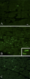
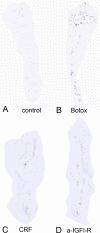
Results: Injection of botulinum toxin into rabbit eyelids resulted in a significant increase in the density of neuromuscular junctions at one and two weeks, and an even greater increase in neuromuscular junction density by four weeks after treatment. Treatment with either CRF or anti-IGFIR completely prevented this increase in neuromuscular junction density.
Conclusions: The return of function after botulinum toxin-induced muscle paralysis is due to terminal sprouting and formation of new neuromuscular junctions within the paralyzed muscles. Injection with CRF or anti-IGFIR after botulinum toxin treatment prevents this sprouting, which in turn should increase the duration of effectiveness of single botulinum toxin treatments. Future physiology studies will address this. Prolonging botulinum toxin's clinical efficacy should decrease the number of injections needed for patient muscle spasm relief, decreasing the risk of negative side effects and changes in drug effectiveness that often occurs over a lifetime of botulinum toxin exposure.
Figures


References
-
- Scott AB, Rosenbaum A, Collins CC. Pharmacologic weakening of extraocular muscles. Invest Ophthalmol. 1973;12:924–927 - PubMed
-
- Dutton JJ, Buckley EG. Long-term results and complications of botulinum A toxin in the treatment of blepharospasm. Ophthalmology. 1988;95:1529–1534 - PubMed
-
- Holds JB, Fogg SG, Anderson RL. Botulinum A toxin injection. Failures in clinical practice and a biomechanical system for the study of toxin-induced paralysis. Ophthal Plast Reconstr Surg. 1990;6:252–259 - PubMed
-
- Jankovic J, Schwartz K. Response and immunoresistance to botulinum toxin injections. Neurology. 1995;45:1743–1746 - PubMed
Publication types
MeSH terms
Substances
Grants and funding
LinkOut - more resources
Full Text Sources
Other Literature Sources
Medical

