Review
. 2010 Dec;83(996):998-1009.
doi: 10.1259/bjr/31200593.

Pneumonia in the immunocompetent patient

Affiliations
Review

Pneumonia in the immunocompetent patient

J H Reynolds et al. Br J Radiol. 2010 Dec.

Abstract

Pneumonia is an acute inflammation of the lower respiratory tract. Lower respiratory tract infection is a major cause of mortality worldwide. Pneumonia is most common at the extremes of life. Predisposing factors in children include an under-developed immune system together with other factors, such as malnutrition and over-crowding. In adults, tobacco smoking is the single most important preventable risk factor. The commonest infecting organisms in children are respiratory viruses and Streptoccocus pneumoniae. In adults, pneumonia can be broadly classified, on the basis of chest radiographic appearance, into lobar pneumonia, bronchopneumonia and pneumonia producing an interstitial pattern. Lobar pneumonia is most commonly associated with community acquired pneumonia, bronchopneumonia with hospital acquired infection and an interstitial pattern with the so called atypical pneumonias, which can be caused by viruses or organisms such as Mycoplasma pneumoniae. Most cases of pneumonia can be managed with chest radiographs as the only form of imaging, but CT can detect pneumonia not visible on the chest radiograph and may be of value, particularly in the hospital setting. Complications of pneumonia include pleural effusion, empyema and lung abscess. The chest radiograph may initially indicate an effusion but ultrasound is more sensitive, allows characterisation in some cases and can guide catheter placement for drainage. CT can also be used to characterise and estimate the extent of pleural disease. Most lung abscesses respond to medical therapy, with surgery and image guided catheter drainage serving as options for those cases who do not respond.


Figures

Figure 1
Figure 1
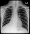
Peripheral right upper lobe consolidation in an adult with community-acquired streptococcal pneumonia.
Figure 2
Figure 2
Chest radiograph demonstrating a foreign body in the right intermediate bronchus.
Figure 3
Figure 3
(a) Chest radiograph and (b) CT scan performed on the same day showing a large left effusion with extensive collapse/consolidation in the left lung on the chest radiograph. The CT scan shows the large left empyema with underlying collapse of the left lung. The lingula shows patchy enhancement with contrast suggestive of infarction.
Figure 4
Figure 4
Chest radiograph illustrating “round pneumonia”, which is seen as a circular opacity adjacent to the right hilum.
Figure 5
Figure 5
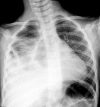
Chest radiograph with extensive collapse/consolidation with a loculated pneumothorax in a child with staphylococcal-Panton–Valentine leukocidin pneumonia.
Figure 6
Figure 6
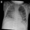
Chest radiograph with extensive bilateral consolidation in a child with staphylococcal-Panton–Valentine leukocidin pneumonia.
Figure 7
Figure 7
(a) Chest radiograph showing left lower zone bronchopneumonia in a hospitalised patient. (b) The corresponding CT image demonstrates bilateral basal consolidation. Note the lack of air bronchograms on both the CT and chest radiograph images.
Figure 8
Figure 8
Chest radiograph of an intensive care patient showing bilateral patchy consolidation owing to a hospital-acquired infection. The presence of pseudomonas was confirmed on sputum culture.
Figure 9
Figure 9
CT image with a centrilobular nodular pattern from a patient with Mycoplasma pneumoniae infection in the right lower lobe.
Figure 10
Figure 10
Chest radiograph showing hyperinflation with bilateral infiltrates in a child with bronchiolitis.
Figure 11
Figure 11
Bilateral infiltrates and ground-glass opacity on the chest radiograph of a child with human metapneumovirus infection.
Figure 12
Figure 12
CT image demonstrating patchy ground-glass opacity in a patient with influenza viral infection with pulmonary involvement.
Figure 13
Figure 13
CT image demonstrating thickening and enhancement of the parietal pleura (black arrowhead) and visceral pleura (white arrowhead) with fluid in between — the so-called “split pleura sign”.
Figure 14
Figure 14
CT image of a lung abscess with a thick, enhancing wall and an air fluid level within (arrow).

Similar articles

Cited by

References

    1. World Health Organization The global burden of disease: 2004 update. WHO Press, 2008
    1. Hirschtick R, Glossroth J, Jordan M, Wilcosky T, Wallace J, Kvale T. Bacterial pneumonia in persons infected with the human immunodeficiency virus. Pulmonary Complications of HIV Infection Study Group. N Engl J Med 1995;333:845–51 - PubMed
    1. Shariatzadeh M, Huang J, Tyrrell G, Johnson M, Marrie T. Bacteremic pneumococcal pneumonia: a prospective study in Edmonton and neighbouring municipalities. Medicine (Baltimore) 2005;84:147–61 - PubMed
    1. Tuomanen E, Austrian R, Masure HR. Pathogenesis of pneumococcal infection. N Engl J Med 2009;332:1280–4 - PubMed
    1. Kin N, Crawford D, Liu J, Behrens T, Kearney J. DNA microarray gene expression profile of marginal zone versus follicular B cells and idiotype positive marginal zone B cells before and after immunization with Streptococcus pneumoniae. J Immunol 2008;180:6663–74 - PMC - PubMed