. 2010 Dec;239(12):3446-66.
doi: 10.1002/dvdy.22484.

Developmental expression patterns of candidate cofactors for vertebrate six family transcription factors

Affiliations

Developmental expression patterns of candidate cofactors for vertebrate six family transcription factors

Karen M Neilson et al. Dev Dyn. 2010 Dec.

Abstract

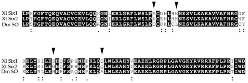
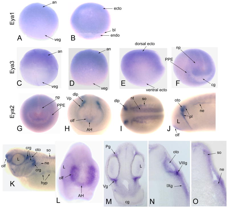
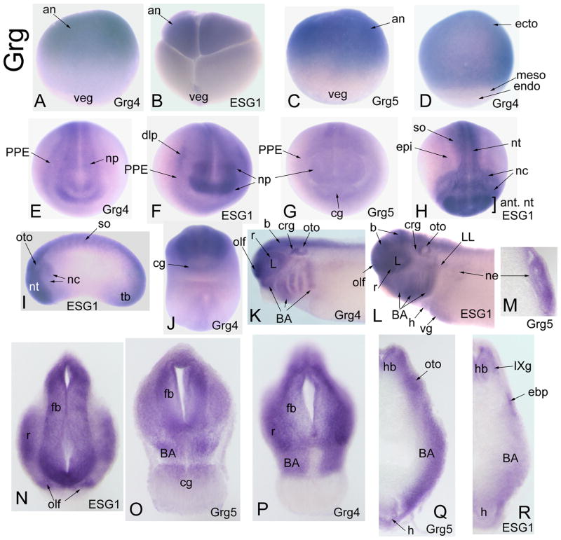
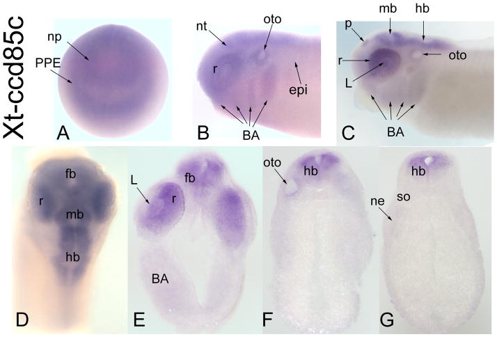
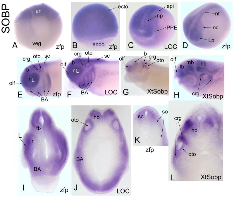
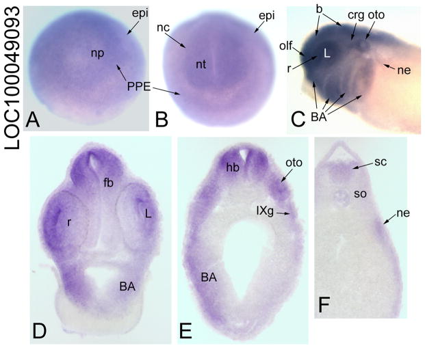
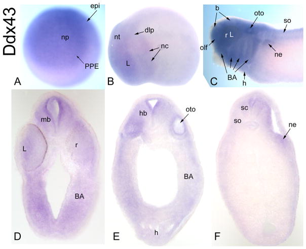
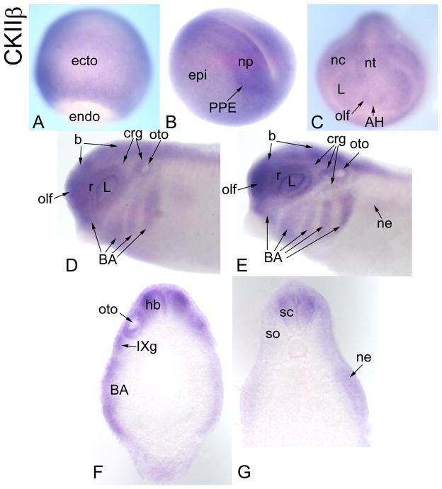
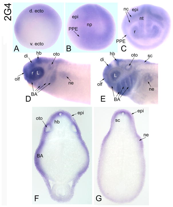
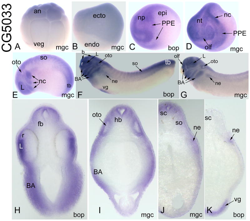
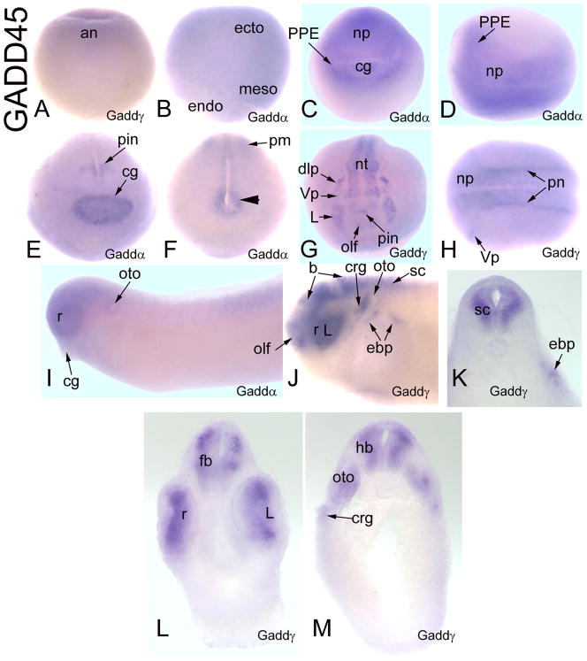
Six family transcription factors play important roles in craniofacial development. Their transcriptional activity can be modified by cofactor proteins. Two Six genes and one cofactor gene (Eya1) are involved in the human Branchio-otic (BO) and Branchio-otic-renal (BOR) syndromes. However, mutations in Six and Eya genes only account for approximately half of these patients. To discover potential new causative genes, we searched the Xenopus genome for orthologues of Drosophila cofactor proteins that interact with the fly Six-related factor, SO. We identified 33 Xenopus genes with high sequence identity to 20 of the 25 fly SO-interacting proteins. We provide the developmental expression patterns of the Xenopus orthologues for 11 of the fly genes, and demonstrate that all are expressed in developing craniofacial tissues with at least partial overlap with Six1/Six2. We speculate that these genes may function as Six-interacting partners with important roles in vertebrate craniofacial development and perhaps congenital syndromes.


Figures

Figure 1
Figure 1
Fly SO, Xenopus Six1 and Xenopus Six2 proteins have a high level of amino acid identity (82.5%; black boxes) throughout their Six domain (SD, aa 1-120), the protein-protein interaction domain. Only 4 amino acid substitutions are non-conserved (arrowheads). All other changes in sequence involve conserved (:) or semi conserved (.) amino acids. The alignment was performed using Clustal W version 2.0 (Larkin et al., 2007). Dm, Drosophila melanogaster; Xl, Xenopus laevis; Xt, Xenopus tropicalis.
Figure 2
Figure 2
Expression of Eya genes. Eya1 is diffusely expressed throughout the ectoderm (ecto) of (A) the animal (an) cap of the blastula and (B) the gastrula [Side views]. veg, vegetal pole; bl, blastopore lip; endo, endoderm. Eya3 is (C) detected in the animal blastomeres of the 32-cell embryo, (D) the animal cap of the blastula, and (E) enhanced in the dorsal ectoderm of the late gastrula [Side views]. (F) At neural plate stages, Eya3 is expressed throughout the neural plate (np) and pre-placodal ectoderm (PPE), but not the cement gland (cg) [Antero-lateral view]. (GO) Eya2 expression. (G) Eya2 is diffusely expressed throughout the neural plate and PPE [Anterior view]. Upon neural tube closure, Eya2 is (H) strongly expressed in the adeno-hypophyseal (AH), olfactory (olf), trigeminal (Vp) and dorso-lateral (dlp) placodes. (I) It also is detected in the caudal neural tube (nt) and somites (so) [H, Anterior view; I, Dorsal view]. At tail bud (J) and larval (K, L) stages, Eya2 is strongly expressed in the olfactory (olf), otic (oto), and various other placodes (pl) and their cranial ganglion derivatives (crg). Note that the lens placode (L) does not express Eya2. There is additional expression in the nephric mesoderm (ne) and hypaxial muscle precursors (hyp) [J, K, Side views; L, anterior view]. In transverse sections at the levels of the diencephalon (M), otocyst (N) and caudal hindbrain (O), Eya2 expression is notable in the cranial ganglia (profundal, Pg; maxillo-mandibular branch of the trigeminal, Vg; VIIIg, IXg), otocyst, somitic and nephric mesoderm.
Figure 3
Figure 3
Expression of Groucho-related genes (Grg). (A) Stage VI oocyte with Grg4 transcripts enhanced on animal (an) side [Side view]. (B) 8-cell embryo with maternal ESG1 transcripts enhanced in the animal blastomeres [Side view]. (C) In the blastula, animal cap ectoderm expresses Grg5 [Side view]. (D) In the gastrula, the entire ectoderm expresses Grg4, whereas the pre-involuted mesoderm (meso) and endoderm do not [Side view]. At neural plate stages, Grg4 (E), ESG1 (F) and Grg5 (G) are expressed in the neural plate and PPE; Grg5 is also expressed in the cement gland [Anterior views]. (H) ESG1 is expressed throughout the neural tube, but most strongly in the anterior part (ant. nt). It is also detected in the neural crest (nc), dorsal epidermis (epi) and somites [Dorsal view]. (I) At tail bud stages, ESG1 expression is strong in the entire neural tube, migrating neural crest, somites, otocyst and tail bud (tb) [Side view]. (J) At tail bud stages, Grg4 is weakly detected in the cement gland [Ventral view]. (K) At larval stages, Grg4 is expressed throughout the brain (b), retina (r), placode derivatives (crg, L, olf, oto) and the branchial arches (BA) [Side view]. (L) ESG1 is expressed in the same tissues, and also is detected in the heart (h), ventral gut (vg), nephric mesoderm and the initial outgrowth of the lateral line (LL) [Side view]. In transverse sections at the levels of the anterior trunk (M), forebrain (N, O, P) and hindbrain (Q, R), expression of the various Grg-related genes is noted in the nephric mesoderm, forebrain (fb), hindbrain (hb), retina, branchial arches, cement gland, various placodes (olf, oto, IXg, epibranchial {ebp}) and heart.
Figure 4
Figure 4
Expression of a CG17265-related gene, Xt-ccd85c. (A) Diffuse expression of Xt-ccd85c throughout the anterior neural plate and PPE [Anterior view]. (B) At neural tube closure, there is diffuse expression throughout the neural tube, retina dorsal epidermis, otocyst and branchial arches [Side view]. (C) At late tail bud stages the pineal (p), retina, midbrain and hindbrain are stained. There also is weak staining in the lens, otocyst and branchial arches [Side view]. (D) Dorsal view at larval stage showing expression extending into the forebrain. Transverse sections at forebrain (E), hindbrain (F) and caudal hindbrain (G) demonstrate restricted neural expression.
Figure 5
Figure 5
Expression of SOBP-related genes. (A) zfp198 mRNA is concentrated in the animal blastomeres at the 8-cell stage [Side view]. (B) zfp198 is expressed throughout the ectoderm of the gastrula [Side view]. (C) At early neural plate, LOC414497 is expressed throughout the neural plate, PPE and dorsal epidermis [Antero-lateral view]. (D) At neural tube closure, neural crest and lens placode (Lp) expression of zfp198 becomes apparent [Antero-lateral view]. (E, F, G) At late tail bud, zfp198 (E) and LOC414497 (F) are expressed throughout the brain, retina, placode derivatives (olf, L, crg, oto) and branchial arches, whereas Xt-Sobp (G) is expressed in patches in the brain and in several placode derivatives (olf, crg, oto) [Side view]. (H) At larval stages, Xt-Sobp expression extends into the forebrain [Side view]. Transverse sections at forebrain (I), hindbrain (J) and spinal (K) levels demonstrate expression of zfg198 and LOC414497 in neural (fb, r, hb, sc), branchial arch, otocyst and somites. (L) Transverse section at hindbrain demonstrates Xt-Sobp more restricted expression in lateral hindbrain, otocyst and cranial ganglia.
Figure 6
Figure 6
Expression of a CG1135-related gene. (A) LOC100049093 is expressed diffusely through the neural plate, PPE and dorsal epidermis (epi) [Anterior view]. (B) At neural tube closure it is slightly enhanced in the neural tube, PPE and neural crest [Anterior view] (C) At larval stages, it is detected in the brain, retina, several placode derivatives (L, olf, oto, crg), branchial arches and nephric mesoderm [Side view]. Transverse sections at forebrain (D), hindbrain (E) and spinal (F) levels demonstrate extensive neural (fb, r, hb, sc), placodal (L, oto, IXg), and branchial arch, and weak expression in the nephric mesoderm.
Figure 7
Figure 7
Expression of a CG7878-related gene. (A) Ddx43 is diffusely expressed throughout the neural plate, PPE and dorsal epidermis [Anterior view]. (B) At neural tube closure, it is very weakly expressed in the neural tube, a few placodes (dlp, L) and migrating neural crest [Side view]. (C) At larval stages it is expressed throughout the brain, retina, several placode derivatives (olf, oto, L), branchial arches, somites, heart and nephric mesoderm [Side view]. Transverse sections at midbrain (D), hindbrain (E) and spinal (F) levels demonstrate neural (mb, r, hb, sc), lens, branchial arch, heart, otocyst, somite and nephric mesoderm expression.
Figure 8
Figure 8
Expression of a CKIIβ-related gene. (A) CKIIβ is expressed throughout the ectoderm of the gastrula [Dorsal view]. (B) At neural plate stages, epidermal staining continues, but expression is enhanced in the neural plate and PPE [Antero-lateral view]. (C) At neural tube closure, CKIIβ is expressed throughout the neural tube, in placodes (AH, olf, L) and neural crest [Anterior view]. (D) At tail bud stages, brain, retina, placodal structures (olf, L, crg, oto) and branchial arches are stained [Side view]. (E) At larval stages, the nephric mesoderm is additionally stained [Side view]. Transverse sections at hindbrain (F) and spinal (G) levels demonstrate neural (hb, sc), branchial arch, otocyst, cranial ganglion (IXg), somite and nephric mesoderm expression.
Figure 9
Figure 9
Expression of a CG10576-related gene. (A) At late gastrula stages, the ectodermal expression of 2G4 is enhanced on the dorsal side (d. ecto) [v. ecto, ventral ectoderm; Side view]. (B) At neural plate stages, 2G4 is expressed diffusely throughout the neural plate, PPE and dorsal epidermis [Antero-lateral view]. (C) 2G4 is expressed throughout the neural tube, retina, PPE, cranial neural crest and epidermis [Anterior view]. (D) At tail bud stages, 2G4 is intensely expressed in the retina, diencephalon (di), hindbrain, placodes (olf, L, oto), branchial arches and weakly in the nephric mesoderm [Side view]. (E) At larval stages, the neural tube expression expands to the dorsal spinal cord, and nephric mesoderm staining is prominent [Side view]. Transverse sections at hindbrain (F) and spinal (G) levels demonstrate neural (hb, sc), branchial arch, otocyst, epidermal and nephric mesoderm expression.
Figure 10
Figure 10
Expression of CG5033-related genes. (A) MGC68939 mRNA is detected in animal blastomeres of the16-cell embryo [Side view]. (B) It is expressed throughout the ectoderm of the gastrula, but not in the endoderm [Side view]. (C) Bop-1 is expressed throughout the neural plate, PPE and dorsal epidermis [Antero-lateral view]. (D) MGC68939 is expressed in the anterior neural tube, cranial neural crest, PPE and some placodes (e.g., olf) [Antero-lateral view]. (E) At early tail bud, MGC68939 is expressed in placodes (L, oto), the migrating neural crest, somites and tail bud [Side view]. (F) At late tail bud, Bop-1 is expressed in the brain, retina, placodes (L, oto), branchial arches, somites, nephric mesoderm, tail bud and ventral gut [Side view]. (G) At late tail bud stages, MGC68939 staining is very similar to that of Bop-1, ventral gut staining is not detected until larval stages [Side view]. Transverse sections at forebrain (H), hindbrain (I), rostral spinal (J) and more caudal spinal (K) levels demonstrate neural (fb, r, hb, sc), branchial arch, lens, otocyst, somite, nephric mesoderm and ventral gut expression.
Figure 11
Figure 11
Expression of Gadd45-related genes. (A) Weak expression of Gadd45γ in the animal cap ectoderm of the blastula [Side view]. (B) Gadd45α is diffusely expressed in the ectoderm and non-involuted mesoderm of the gastrula [Side view]. (C) and (D) Gadd45α is expressed throughout the neural plate, PPE and cement gland [C, Anterior view; D, Dorsal view]. At neural tube closure, Gadd45α expression in the anterior part of the embryo (E) is enhanced in the cement gland and pineal, and in the posterior part (F) in a patch of paraxial mesoderm (pm) and in mesoderm surrounding the cloaca (arrowhead). (G) At neural tube stages, Gadd45γ is expressed in the neural tube, including pineal, and multiple placodes (olf, L, Vp, dlp) [Anterior view]. (H) At neural plate Gadd45γ is expressed in stripes of primary neurons (pn) and the trigeminal placode (Vp) [Dorsal view]. (I) At larval stages, Gadd45α expression becomes weak and diffuse throughout the head and anterior trunk, and is lost from the cement gland. [Side view]. (J) At larval stages, Gadd45γ expression is confined to the brain, retina, anterior spinal cord and placode derivatives (olf, L, crg, oto, ebp) [Side view]. Transverse sections at forebrain (L), hindbrain (M), rostral spinal (K) levels demonstrate neural (fb, r, hb, sc) and placodal (L, oto, crg, ebp) expression.

Similar articles

Cited by

References

    1. Abe Y, Oka A, Mizuguchi M, Igarashi T, Ishikawa S, Aburatani H, Yokoyama S, Asahara H, Nagao K, Yamada M, Miyashita T. Eya4, deleted in a case with middle interhemispheric variant of Holoprosencephaly interacts with Six3 both physically and functionally. Human Mutation. 2009;30:946–955. - PubMed
    1. Abdelhak S, Kalatzis V, Heilig R, Compain S, Samson D, Vincent C, Weil D, Cruaud C, Sahly I, Leibovici M, Bitner-Glindzicz M, Francis M, Lacombe D, Vigneron J, Charachon R, Boven K, Bedbeder P, Van Regemorter N, Weissenbach J, Petit C. A human homologue of the Drosophila eyes absent gene underlies branchio-oto-renal (BOR) syndrome and identifies a novel gene family. Nat Genet. 1997;15:157–164. - PubMed
    1. Bai J, Montell D. Eyes absent, a key repressor of polar cell fate during Drosophila oogenesis. Development. 2002;129:5377–5388. - PubMed
    1. Baker CV, Bronner-Fraser M. Vertebrate cranial placodes. I. Embryonic induction. Dev Biol. 2001;232:1–61. - PubMed
    1. Bernard OA, Bisson-LeConiat M, Ballerini P, Mauchauffe M, Della Valle V, Monni R, Nguyen Khac F, Mercher T, Penard-Lacronique V, Paturaud P, Gressin L, Hellig Rl, Daniel MT, Lessard M, Berger R. A new recurrent and specific cryptic translocation, t(5;14)(q35;q32), is associated with expression of the Hox11L2 gene in T acute lymphoblastic leukemia. Leukemia. 2001;15:1495–1504. - PubMed

Publication types

MeSH terms