. 2010 Oct;37(10):5306-12.
doi: 10.1118/1.3491806.

Prospective-gated cardiac micro-CT imaging of free-breathing mice using carbon nanotube field emission x-ray

Affiliations

Prospective-gated cardiac micro-CT imaging of free-breathing mice using carbon nanotube field emission x-ray

Guohua Cao et al. Med Phys. 2010 Oct.

Abstract

Purpose: Carbon nanotube (CNT) based field emission x-ray source technology has recently been investigated for diagnostic imaging applications because of its attractive characteristics including electronic programmability, fast switching, distributed source, and multiplexing. The purpose of this article is to demonstrate the potential of this technology for high-resolution prospective-gated cardiac micro-CT imaging.

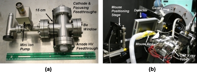
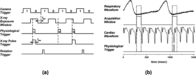
Methods: A dynamic cone-beam micro-CT scanner was constructed using a rotating gantry, a stationary mouse bed, a flat-panel detector, and a sealed CNT based microfocus x-ray source. The compact single-beam CNT x-ray source was operated at 50 KVp and 2 mA anode current with 100 microm x 100 microm effective focal spot size. Using an intravenously administered iodinated blood-pool contrast agent, prospective cardiac and respiratory-gated micro-CT images of beating mouse hearts were obtained from ten anesthetized free-breathing mice in their natural position. Four-dimensional cardiac images were also obtained by gating the image acquisition to different phases in the cardiac cycle.

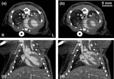
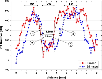
Results: High-resolution CT images of beating mouse hearts were obtained at 15 ms temporal resolution and 6.2 lp/mm spatial resolution at 10% of system MTF. The images were reconstructed at 76 microm isotropic voxel size. The data acquisition time for two cardiac phases was 44 +/- 9 min. The CT values observed within the ventricles and the ventricle wall were 455 +/- 49 and 120 +/- 48 HU, respectively. The entrance dose for the acquisition of a single phase of the cardiac cycle was 0.10 Gy.

Conclusions: A high-resolution dynamic micro-CT scanner was developed from a compact CNT microfocus x-ray source and its feasibility for prospective-gated cardiac micro-CT imaging of free-breathing mice under their natural position was demonstrated.


Figures

Figure 1
Figure 1
Pictures of (a) the compact CNT field emission microfocus x-ray tube and (b) the desktop in vivo micro-CT scanner that consists of the CNT field emission microfocus x-ray tube, a flat-panel detector, a small-diameter rotating gantry, and a stationary mouse bed. The x-ray tube’s body dimension is 150 mm×70 mm×70 mm.
Figure 2
Figure 2
(a) Illustrative timing diagram for the dynamic gating method that the micro-CT system used to gate the x-ray exposure and image acquisition to a nonperiodic physiological trigger signal. The camera readout (470 ms) and integration (500 ms) regions are designated as 1 and 0, respectively. tpw is the temporal width of an x-ray pulse (15 ms for cardiac imaging). Three nonequally spaced physiological triggers (labeled as 1, 2, and 3) are used to illustrate the dynamic gating. (b) Generation of the physiological trigger corresponding to the R peak in the ECG cycle and end-expiration in the respiration cycle. The acquisition window was 100 ms in duration and defined during end-expiration of the respiration cycle. A physiological trigger was generated if an R wave occurred within the acquisition window. Physiological triggers corresponding to other points in the ECG cycle were generated by adding a constant delay after detecting the R peak. A relatively constant heart rate (±10%) was maintained throughout each scan.
Figure 3
Figure 3
[(a) and (b)] Axial and [(c) and (d)] coronal slice images of a C57BL∕6 mouse at [(a) and (c)] 0 and [(b) and (d)] 55 ms after the R wave. All images have the same display window and level. The scanning parameters were 50 kVp, 2 mA, 15 ms pulse width, and 400 views over 200° at a step angle of 0.5°. These images represent 76 μm sections taken at the same slice locations from the 3D volumes reconstructed at each time point. The voxel spacing in-plane is also 76 μm. The major anatomic structures of the cardiopulmonary vascular system are readily identified in the contrast enhanced images. The AO, LV, and RV are labeled for reference.
Figure 4
Figure 4
Intensity profiles along the two lines within the axial images shown in Figs. 3a, 3b. For each intensity profile, the two boundary regions between the ventricles and the ventricle wall were performed with a linear fit. The fitting slopes are shown in Table 1. The three (RV, VW, and LV) sections of the line profiles are labeled in the plot. The width at the midheight of the VW section changed from 1.3 mm at 0 ms to 1.8 mm at 55 ms, representing a change of 0.5 mm in the ventricle wall thickness from diastole to systole.

Similar articles

Cited by

References

    1. Badea C. T., Bucholz E., Hedlund L. W., Rockman H. A., and Johnson G. A., “Imaging methods for morphological and functional phenotyping of the rodent heart,” Toxicol. Pathol. TOPADD 34, 111–117 (2006).10.1080/01926230500404126 - DOI - PubMed
    1. Johnson K., “Introduction to rodent cardiac imaging,” ILAR J. IJLOAC 49, 27–34 (2008). - PubMed
    1. Sera T., Yokota H., Fujisaki K., Fukasaku K., Tachibana H., Uesugi K., Yagi N., and Himeno R., “Development of high-resolution 4D in vivo-CT for visualization of cardiac and respiratory deformations of small animals,” Phys. Med. Biol. PHMBA7 53, 4285–4301 (2008).10.1088/0031-9155/53/16/005 - DOI - PubMed
    1. Badea C. T., Fubara B., Hedlund L. W., and Johnson G. A., “4-D micro-CT of the mouse heart,” Mol. Imaging MIOMBP 4, 110–116 (2005). - PubMed
    1. Drangova M., Ford N. L., Detombe S. A., Wheatley A. R., and Holdsworth D. W., “Fast retrospectively gated quantitative four-dimensional (4D) cardiac micro computed tomography imaging of free-breathing mice,” Invest. Radiol. INVRAV 42, 85–94 (2007).10.1097/01.rli.0000251572.56139.a3 - DOI - PubMed

Publication types

MeSH terms

Substances