

In vivo imaging of corneal inflammation: new tools for clinical practice and research
- PMID: 21090997
- PMCID: PMC3146960
- DOI: 10.3109/08820538.2010.518542
In vivo imaging of corneal inflammation: new tools for clinical practice and research
Abstract
Purpose: Infectious and inflammatory corneal diseases are a major cause of blindness. To date, assessment of corneal inflammation, has only been possible by slit-lamp biomicroscopy. The purpose of this study is to review the current state of imaging technologies enabling in vivo imaging of inflammation in the cornea.
Methods: Literature review of peer-reviewed articles on in vivo imaging modalities.
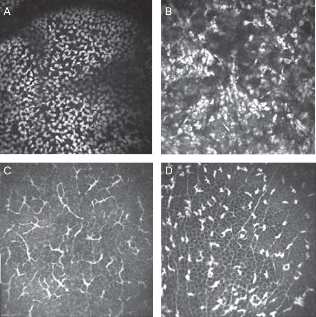
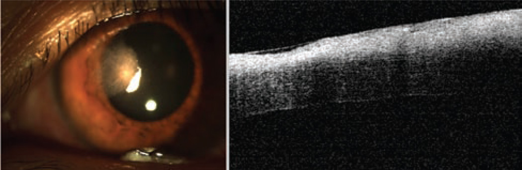
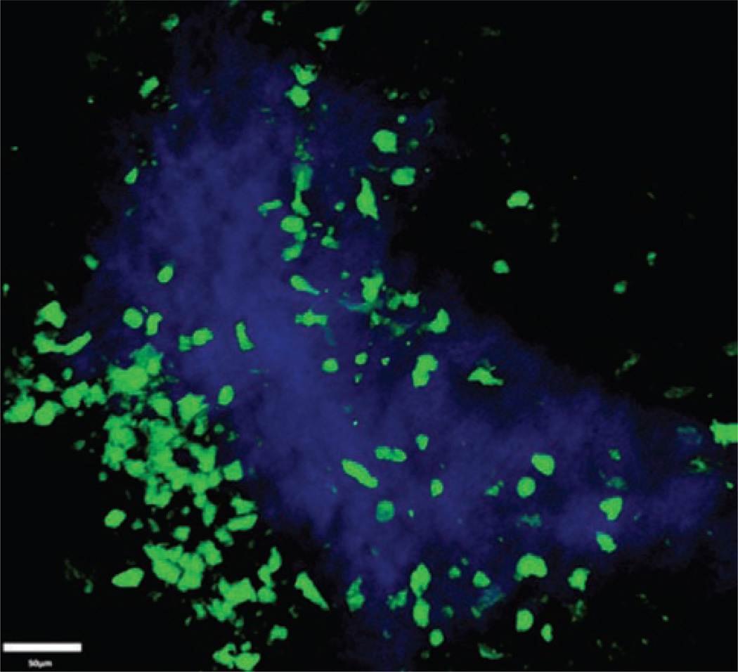
Results: Current means of diagnosis and treatment follow-up for immune and infectious keratitis are limited to slit-lamp biomicroscopy. Several modalities are currently emerging, allowing for in vivo imaging of corneal inflammation, including in vivo confocal microscopy, anterior segment optical coherence tomography, and intravital multiphoton microscopy.
Conclusion: Several in vivo imaging technologies are currently evolving, allowing for objective assessment of corneal inflammation and treatment response.
Conflict of interest statement
Figures
Similar articles
-
[Quantitative evaluation of infectious keratitis by anterior segment optical coherence tomography].J Fr Ophtalmol. 2020 Oct;43(8):731-741. doi: 10.1016/j.jfo.2019.11.024. Epub 2020 Jul 1. J Fr Ophtalmol. 2020. PMID: 32622634 French.
-
Corneal assessment technologies: current status.Surv Ophthalmol. 2014 Nov-Dec;59(6):599-614. doi: 10.1016/j.survophthal.2014.05.001. Epub 2014 May 24. Surv Ophthalmol. 2014. PMID: 25223496 Review.
-
In vivo imaging of coin-shaped lesions in cytomegalovirus corneal endotheliitis by anterior segment optical coherence tomography.Cornea. 2014 Dec;33(12):1332-5. doi: 10.1097/ICO.0000000000000269. Cornea. 2014. PMID: 25285587
-
Mulitmodal Corneal Imaging of Genetically Confirmed Keratitis-Ichthyosis-Deafness Syndrome.Cornea. 2020 Nov;39(11):1446-1449. doi: 10.1097/ICO.0000000000002415. Cornea. 2020. PMID: 32618851
-
Recent advances in corneal neovascularization imaging.Exp Eye Res. 2024 Jul;244:109930. doi: 10.1016/j.exer.2024.109930. Epub 2024 May 16. Exp Eye Res. 2024. PMID: 38750782 Review.
Cited by
-
Corneal Innervation and Sensation: The Eye and Beyond.Yale J Biol Med. 2018 Mar 28;91(1):13-21. eCollection 2018 Mar. Yale J Biol Med. 2018. PMID: 29599653 Free PMC article. Review.
-
Comparing in-vivo confocal microscopy and ex-vivo light and scanning electron microscopy images of the hairs of the pine processionary caterpillar embedded in the cornea: Report of three cases.Indian J Ophthalmol. 2020 Aug;68(8):1672-1675. doi: 10.4103/ijo.IJO_1735_19. Indian J Ophthalmol. 2020. PMID: 32709819 Free PMC article.
-
Degeneration and Regeneration of Subbasal Corneal Nerves after Infectious Keratitis: A Longitudinal In Vivo Confocal Microscopy Study.Ophthalmology. 2015 Nov;122(11):2200-9. doi: 10.1016/j.ophtha.2015.06.047. Epub 2015 Aug 6. Ophthalmology. 2015. PMID: 26256833 Free PMC article. Clinical Trial.
-
Comparison of reflectance confocal microscopy and two-photon second harmonic generation microscopy in fungal keratitis rabbit model ex vivo.Biomed Opt Express. 2016 Jan 26;7(2):677-87. doi: 10.1364/BOE.7.000677. eCollection 2016 Feb 1. Biomed Opt Express. 2016. PMID: 26977371 Free PMC article.
-
What's new in dry eye disease diagnosis? Current advances and challenges.F1000Res. 2018 Dec 19;7:F1000 Faculty Rev-1952. doi: 10.12688/f1000research.16468.1. eCollection 2018. F1000Res. 2018. PMID: 30613376 Free PMC article. Review.
References
-
- Keay L, Edwards K, Naduvilath T, et al. Microbial keratitis predisposing factors and morbidity. Ophthalmology. 2006 Jan;113(1):109–116. - PubMed
-
- McLeod SD, LaBree LD, Tayyanipour R, et al. The importance of initial management in the treatment of severe infectious corneal ulcers. Ophthalmology. 1995 Dec;102(12):1943–1948. - PubMed
-
- Swanson EA, Izatt JA, Hee MR, et al. In vivo retinal imaging by optical coherence tomography. Opt Lett. 1993 Nov 1;18(21):1864–1866. - PubMed
Publication types
MeSH terms
Grants and funding
LinkOut - more resources
Full Text Sources