

In vivo imaging of corneal inflammation: new tools for clinical practice and research
- PMID: 21090997
- PMCID: PMC3146960
- DOI: 10.3109/08820538.2010.518542
In vivo imaging of corneal inflammation: new tools for clinical practice and research
Abstract
Purpose: Infectious and inflammatory corneal diseases are a major cause of blindness. To date, assessment of corneal inflammation, has only been possible by slit-lamp biomicroscopy. The purpose of this study is to review the current state of imaging technologies enabling in vivo imaging of inflammation in the cornea.
Methods: Literature review of peer-reviewed articles on in vivo imaging modalities.
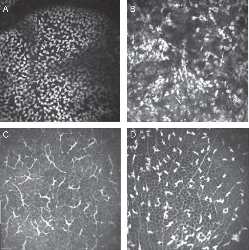
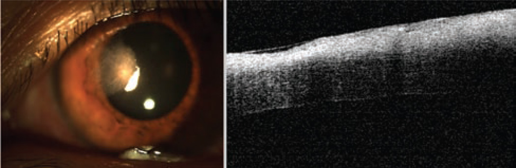
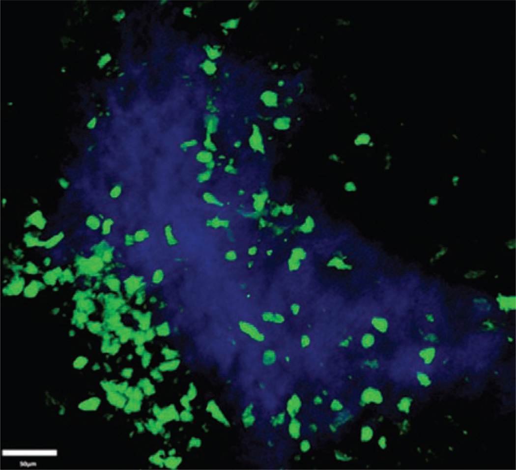
Results: Current means of diagnosis and treatment follow-up for immune and infectious keratitis are limited to slit-lamp biomicroscopy. Several modalities are currently emerging, allowing for in vivo imaging of corneal inflammation, including in vivo confocal microscopy, anterior segment optical coherence tomography, and intravital multiphoton microscopy.
Conclusion: Several in vivo imaging technologies are currently evolving, allowing for objective assessment of corneal inflammation and treatment response.
Conflict of interest statement
Figures
References
-
- Keay L, Edwards K, Naduvilath T, et al. Microbial keratitis predisposing factors and morbidity. Ophthalmology. 2006 Jan;113(1):109–116. - PubMed
-
- McLeod SD, LaBree LD, Tayyanipour R, et al. The importance of initial management in the treatment of severe infectious corneal ulcers. Ophthalmology. 1995 Dec;102(12):1943–1948. - PubMed
-
- Swanson EA, Izatt JA, Hee MR, et al. In vivo retinal imaging by optical coherence tomography. Opt Lett. 1993 Nov 1;18(21):1864–1866. - PubMed
Publication types
MeSH terms
Grants and funding
LinkOut - more resources
Full Text Sources