Review
. 2011 Oct 1;15(7):1945-56.
doi: 10.1089/ars.2010.3742. Epub 2011 Mar 23.

Radiation as a risk factor for cardiovascular disease

Affiliations
Review

Radiation as a risk factor for cardiovascular disease

John E Baker et al. Antioxid Redox Signal. .

Abstract

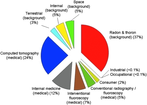
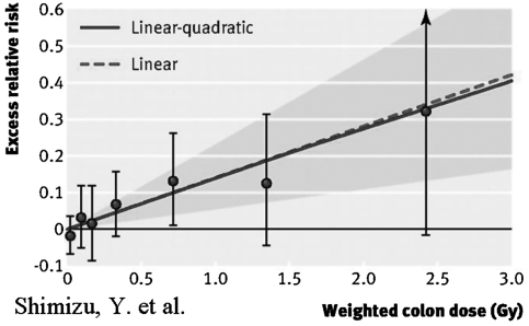
Abstract population are ubiquitous background radiation and medical exposure of patients. From the early 1980s to 2006, the average dose per individual in the United States for all sources of radiation increased by a factor of 1.7-6.2 mSv, with this increase due to the growth of medical imaging procedures. Radiation can place individuals at an increased risk of developing cardiovascular disease. Excess risk of cardiovascular disease occurs a long time after exposure to lower doses of radiation as demonstrated in Japanese atomic bomb survivors. This review examines sources of radiation (atomic bombs, radiation accidents, radiological terrorism, cancer treatment, space exploration, radiosurgery for cardiac arrhythmia, and computed tomography) and the risk for developing cardiovascular disease. The evidence presented suggests an association between cardiovascular disease and exposure to low-to-moderate levels of radiation, as well as the well-known association at high doses. Studies are needed to define the extent that diagnostic and therapeutic radiation results in increased risk factors for cardiovascular disease, to understand the mechanisms involved, and to develop strategies to mitigate or treat radiation-induced cardiovascular disease.


Figures

FIG. 1.
FIG. 1.
Percent contribution of various sources of exposure to the total effective dose per individual in the U.S. population (6.2 mSv) for 2006. Reprinted with permission from the National Council on Radiation Protection and Measurements, http://NCRPonline.ord.
FIG. 2.
FIG. 2.
Radiation dose–response relation (excess relative risk) for death from heart disease, showing linear and linear-quadratic functions. Shaded area is 95% confidence region for fitted linear line. Vertical lines are 95% confidence intervals for specific dose category risks. Point estimates of risk for each dose category are indicated by circles. Reproduced from Shimizu et al. (79) with permission from BMJ Publishing Group Ltd.
FIG. 3.
FIG. 3.
Time-related changes in total cholesterol, low-density lipoprotein (LDL) cholesterol, and triglycerides after total body irradiation (TBI). Data shown as mean ± SD, n = 6/group. *p < 0.05, 10 gray (Gy) versus unirradiated controls. Reproduced from Baker et al. (6) with permission from Taylor & Francis, Ltd. (www.tandf.co.uk/journals).
FIG. 4.
FIG. 4.
Computed tomography reconstruction of coronary arteries at 120 days after TBI, compared to age-matched control. TBI reduced the number of vessels of smaller diameter (<50 μm diameter) but not the epicardial vessels. Reproduced from Baker et al. (6) with permission from Taylor & Francis, Ltd. (www.tandf.co.uk/journals).
FIG. 5.
FIG. 5.
Morphological changes to the coronary vasculature at 120 days after 10 Gy TBI. (A) Heart sections, stained with H&E, show vessel lumen completely blocked (→) as a result of myointimal proliferation 120 days after 10 Gy TBI. The lumen of a comparable vessel in an age-matched unirradiated heart is patent and contains red blood cells (⇒). (B) Heart section, stained with Trichrome, showing increased peri-arterial fibrosis in small caliber coronary vessel 120 days after 10 Gy TBI compared with a comparable vessel in an age-matched control. Fibrosis appears as blue using trichrome staining. Reproduced from Baker et al. (6) with permission from Taylor & Francis, Ltd. (www.tandf.co.uk/journals).
FIG. 6.
FIG. 6.
Change in ventricular function at 120 days after 10 Gy TBI compared with age-matched controls. *p < 0.05 for 10 Gy versus unirradiated age-matched control. Reproduced from Baker et al. (6) with permission from Taylor & Francis, Ltd. (www.tandf.co.uk/journals).

References

    1. These references have been deleted.
    1. Adams MJ. Lipshultz SE. Schwartz C. Fajardo LF. Coen V. Constine LS. Radiation-associated cardiovascular disease: manifestations and management. Semin Radiat Oncol. 2003;13:346–356. - PubMed
    1. Alpen EL. Powers-Risius P. Curtis SB. DeGuzman R. Tumorigenic potential of high-Z, high-LET charged-particle radiations. Radiat Res. 1993;136:382–391. - PubMed
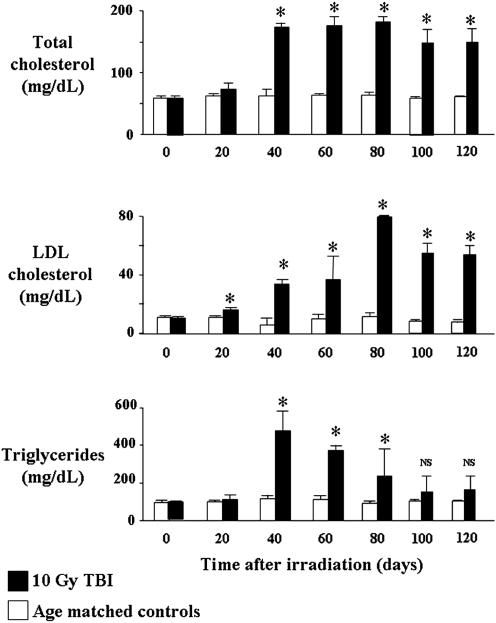
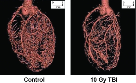
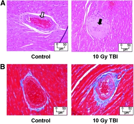
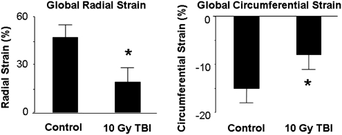
    1. Baker JE. Fish B. Su J. Haworth S. Strande J. Komorowski R. Migrino R. Doppalapudi A. Harmann L. Li X. Hopewell J. Moulder J. 10 Gy Total body irradiation increases risk of coronary sclerosis, degeneration of heart structure and function in a rat model. Int J Radiat Biol. 2009;85:1089–1100. - PMC - PubMed
    1. Barrett-Connor E. Sex differences in coronary heart disease. Why are women so superior? The 1995 Ancel Keys Lecture. Circulation. 1997;95:252–264. - PubMed

Publication types

MeSH terms

LinkOut - more resources