. 2011 Jul;22(7):829-41.
doi: 10.1089/hum.2010.162. Epub 2011 Mar 30.

Genetically modified donor leukocyte transfusion and graft-versus-leukemia effect after allogeneic stem cell transplantation

Affiliations

Genetically modified donor leukocyte transfusion and graft-versus-leukemia effect after allogeneic stem cell transplantation

Sylvia Borchers et al. Hum Gene Ther. 2011 Jul.

Abstract

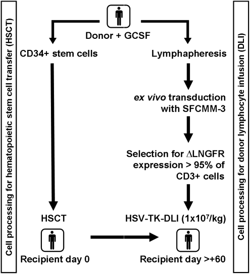
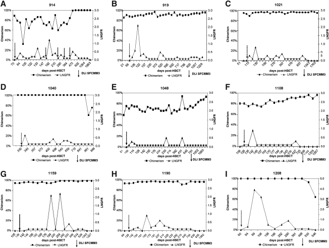
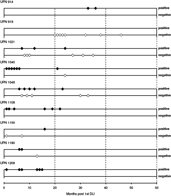
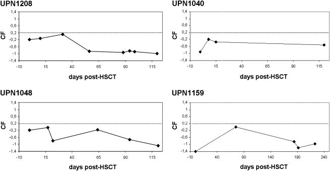
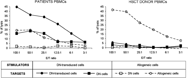
Seven patients with acute myeloid leukemia (AML) and two patients with chronic myelogenous leukemia (CML) were transplanted from HLA-identical sibling donors with CD34(+) cell-enriched stem cells (HSCTs) without further immunosuppression. The myeloablative standard transplantation protocol was adapted to include transfusion of gene-modified donor T cells after HSCT. Donor T cells were transduced with the replication-deficient retrovirus SFCMM-3, which expresses herpes simplex thymidine kinase (HSV-Tk) and a truncated version of low-affinity nerve growth factor receptor (ΔLNGFR) for selection and characterization of transduced cells. Transduced T cells were detectable in all patients during follow-up for up to 5 years after transfusion. Proteomic screening for development of acute graft-versus-host disease (aGvHD) was applied to five of the seven patients with AML. No positivity for the aGvHD grade II-specific proteomic pattern was observed. Only one patient developed aGvHD grade I. To date, three of the patients with AML relapsed; one responded to three escalating transfusions of lymphocytes from the original donor and is in complete remission. Two were retransplanted with non-T cell-depleted peripheral blood stem cells from their original donors and died after retransplantation of septic complications or relapse, respectively. In one patient with CML, loss of bcr-abl gene expression was observed after an expansion of transduced cells. Seven of nine patients are alive and in complete remission.


Figures

FIG. 1.
FIG. 1.
Clinical trial: HSV-TK-DLI. The clinical protocol was designed to include SFCMM-3-transduced donor lymphocyte infusion (DLI) in a myeloablative conditioning regimen and CD34+ cell-enriched hematopoietic stem cell transplantation (HSCT). The donor underwent leukapheresis once either before or at least 6 weeks after granulocyte colony-stimulating factor (G-CSF) administration. In a separate leukapheresis, peripheral blood stem cells were collected and transplanted to the recipient on day 0. Transduction of the cells and safety testing were performed at MolMed (Milan, Italy), and cells were shipped via courier to Hannover. Transfer of the transduced cells was scheduled on day +60 after HSCT. ΔLNGFR, truncated version of low-affinity nerve growth factor receptor; HSV-Tk, herpes simplex virus thymidine kinase.
FIG. 2.
FIG. 2.
Follow-up of transduced donor T cells: (A) UPN 914; (B) UPN 919; (C) UPN 1021; (D) UPN 1040; (E) UPN 1048; (F) UPN 1108; (G) UPN 1159; (H) UPN 1190; (I) UPN 1208. Shown is the percentage of donor chimerism in peripheral blood (solid squares; left y axis) and the percentage of ΔLNGFR-expressing cells (FACS) (triangles; right y axis) plotted against days after HSCT. The time of transfusion of donor lymphocytes is indicated by an arrow (DLI SFCMM-3). UPN 914 received more than one DLI, but only the first one was transduced; all other patients have received only one DLI to date (DLI SFCMM-3). UPN 919 (B) and UPN 1108 (F) received only 4 × 106 SFCMM-3-transduced cells/kg (Table 2). HSCT, hematopoietic stem cell transplantation; LNGFR, low-affinity nerve growth factor receptor.
FIG. 3.
FIG. 3.
T cell receptor repertoire screening. Donor (after transduction and enrichment for LNGFR expression) and patient T cells were isolated, RNA and cDNA were prepared and spectratyping was performed as described in Patients and Methods. Data for all donors and recipients are shown in Supplementary Table S1. Fourteen bifamily primer sets were used to analyze the Vβ family TCR repertoire for each donor and the corresponding patient. Here results are shown for one donor (for patient UPN 1280). Multiplex primers used are indicated: 1, Vβ1 and Vβ12; 2, Vβ13 and Vβ3; 3, Vβ5.1 and Vβ1; 4, Vβ4 and Vβ5.3; 5, Vβ8 and Vβ7; 6, Vβ9 and Vβ14; 7, Vβ11 and Vβ20; 8, Vβ16 and Vβ22; 9, Vβ18 and Vβ23; 10, Vβ17 and Vβ15; 11,Vβ24 and Vβ22; 12, Vβ6.1; 13, Vβ6.2; 14, Vβ12 (single).
FIG. 4.
FIG. 4.
Long-term follow-up of transgene expression in all patients. PCR analyses were performed routinely for all patients in order to ensure transgene expression. To date, six of nine patients have shown HSV-Tk and ΔLNGFR gene expression, three experienced relapse and had no transgene expression at that time, and one (UPN 919) lost expression of the transgenes at about 20 months post-DLI. The x axis of the plot shows months posttransfusion of SFCMM-3 DLI, and positivity or negativity for HSV-Tk gene expression is indicated by diamonds on the y axis in the positive (+) or negative (−) direction.
FIG. 5.
FIG. 5.
Proteomic screening of urine collected post-HSCT. The days post-HSCT (x axis) are plotted against the classification factor (CF) (y axis) as described (Weissinger et al., 2007b). The cutoff for diagnosis of acute GvHD was set to CF = +0.2 and is indicated as a dotted line. Proteomic screening data were obtained from five of nine patients, and four examples are shown here (UPN 1040, 1048, 1159, and 1208).
FIG. 6.
FIG. 6.
Detection of an immune response to HSV-Tk in patient UPN 919. After coculture with SFCMM-3-transduced donor cells (straight line) or allogeneic cells (dashed line), patient and donor PBMCs were tested against the same stimulators and untransduced donor cells in a standard 51Cr release assay. Although patient effectors showed a weaker response to allogeneic targets as compared with donor effectors, only patient T cells showed a specific lytic response to transduced lymphocytes.

References

    1. Bahceci E. Epperson D. Douek D.C., et al. Early reconstitution of the T-cell repertoire after non-myeloablative peripheral blood stem cell transplantation is from post-thymic T-cell expansion and is unaffected by graft-versus-host disease or mixed chimaerism. Br. J. Haematol. 2003;122:934–943. - PubMed
    1. Barrett A.J. Mavroudis D. Tisdale J., et al. T cell-depleted bone marrow transplantation and delayed T cell add-back to control acute GVHD and conserve a graft-versus-leukemia effect. Bone Marrow Transplant. 1998;21:543–551. - PubMed
    1. Bondanza A. Valtolina V. Magnani Z., et al. Suicide gene therapy of graft-versus-host disease induced by central memory human T lymphocytes. Blood. 2006;107:1828–1836. - PubMed
    1. Bonini C. Ciceri F. Marktel S., et al. Suicide-gene-transduced T-cells for the regulation of the graft-versus-leukemia effect. Vox Sanguinis. 1998;74(Suppl. 2):341–343. - PubMed
    1. Briones J. Urbano-Ispizua A. Lawler M., et al. High frequency of donor chimerism after allogeneic transplantation of CD34+-selected peripheral blood cells. Exp. Hematol. 1998;26:415–420. - PubMed

Publication types

MeSH terms

Substances