Comparative Study
. 2011 Feb;28(2):245-57.
doi: 10.1089/neu.2010.1639. Epub 2011 Jan 27.

Differential effects of injury severity on cognition and cellular pathology after contusive brain trauma in the immature rat

Affiliations
Comparative Study

Differential effects of injury severity on cognition and cellular pathology after contusive brain trauma in the immature rat

Jimmy W Huh et al. J Neurotrauma. 2011 Feb.

Abstract

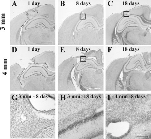
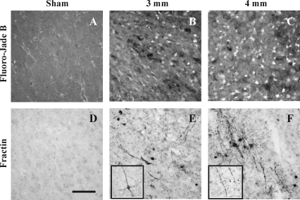
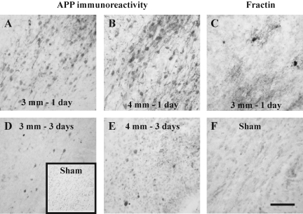
Although diffuse brain damage has been suggested to be the predominant predictor of neurological morbidity following closed head injury in infants and children, the presence of contusions also predicts long-term neurobehavioral dysfunction. Contusive brain trauma in the 17-day-old rat resulted in neurodegeneration and caspase activation in the cortex at 1 day, and in the thalamus at 3 days post-injury, and to a greater extent following a deeper impact. Cortical tissue loss in the 4-mm impact group was significantly greater than that in the 3-mm impact group (p < 0.05), and exhibited a time-dependent increase over the first 3 weeks post-injury. Traumatic axonal injury was observed in the white matter tracts below the site of impact at 1 day, and in the corpus callosum at 3 days, to a greater extent following 4-mm impact. In contrast, cellular caspase-3 activation in these white matter tracts was only observed at 24 h post-injury and was not affected by impact depth. Similarly, neurodegeneration and caspase activation in the hippocampus was restricted to the dentate gyrus and occurred to a similar extent in both injured groups. Only the 4-mm impact group exhibited learning deficits in the first week (p < 0.0001) that was sustained until the third week post-injury (p < 0.0001), while deficits in the 3-mm impact group were seen only at 3 weeks post-injury (p < 0.02). These observations demonstrate that increasing severity of injury in immature animals does not uniformly increase the extent of cellular damage, and that the progression of tissue damage and behavioral deficits varies as a function of injury severity.


Figures

FIG. 1.
FIG. 1.
Cortical tissue loss following contusive brain trauma in 17-day-old rats. Representative images of cresyl violet-cyanine R-stained tissue sections from animals subjected to 3-mm (AC, G, and H), or 4-mm depth of impact (DF, and I), demonstrating progressive loss of cortical tissue after impact. Note hemorrhage and tissue tears on day 1 (A and D) and day 8 (B and E), and pronounced cavitation on days 1 (D), 8 (E), and 18 (F) after 4-mm depth of impact; there was no overt cavitation on day 18 following 3-mm depth of impact (C). Boxes in panels B, C, and E represent areas illustrated at higher magnification in panels G, H, and I, respectively. Note the presence of non-neuronal cells in the gray-white matter interface at both 3-mm (G and H) and 4-mm depths of impact (I). The graph in J demonstrates the quantitative analysis of cortical tissue loss in the injured hemisphere at 8 and 18 days following either 3-mm or 4-mm depth of impact (*p < 0.0002 compared to 8 days after 4-mm depth of impact; #p < 0.0002 compared to the 3-mm group; scale bar in A–F = 1 mm, and for GI = 100 μm).
FIG. 1.
FIG. 1.
Cortical tissue loss following contusive brain trauma in 17-day-old rats. Representative images of cresyl violet-cyanine R-stained tissue sections from animals subjected to 3-mm (AC, G, and H), or 4-mm depth of impact (DF, and I), demonstrating progressive loss of cortical tissue after impact. Note hemorrhage and tissue tears on day 1 (A and D) and day 8 (B and E), and pronounced cavitation on days 1 (D), 8 (E), and 18 (F) after 4-mm depth of impact; there was no overt cavitation on day 18 following 3-mm depth of impact (C). Boxes in panels B, C, and E represent areas illustrated at higher magnification in panels G, H, and I, respectively. Note the presence of non-neuronal cells in the gray-white matter interface at both 3-mm (G and H) and 4-mm depths of impact (I). The graph in J demonstrates the quantitative analysis of cortical tissue loss in the injured hemisphere at 8 and 18 days following either 3-mm or 4-mm depth of impact (*p < 0.0002 compared to 8 days after 4-mm depth of impact; #p < 0.0002 compared to the 3-mm group; scale bar in A–F = 1 mm, and for GI = 100 μm).
FIG. 2.
FIG. 2.
Neurodegeneration and caspase activation in the cortex at 24 h following contusive brain trauma in 17-day-old rats. Photomicrographs represent Fluoro-Jade B reactivity in the cortex adjacent to the site of maximal lesion following 3-mm (B) or 4-mm depth of impact (C). Similar regions in the cortices of sham-injured animals did not contain either Fluoro-Jade B-positive (A) or fractin-labeled neurons (D). In sections adjacent to those used for Fluoro-Jade B staining, fractin immunoreactivity was observed in cell bodies and neurites at both 3-mm (E) and 4-mm depth of impact (F).The inset in panel E demonstrates an example of a fractin-positive neuron that does not show neurite beading; these were occasionally seen within and around the site of maximal lesion. The inset in panel F illustrates a beaded neurite containing fractin. Scale bar in panel D represents 50 μm for AF, and 25 μm for the insets in panels E and F.
FIG. 3.
FIG. 3.
Neurodegeneration and caspase activation in the thalamus following contusive brain trauma in 17-day-old rats. Photomicrographs represent Fluoro-Jade B reactivity in the thalamus at 1 day following 4-mm depth of impact (A), 3 days following either 3-mm (B) or 4-mm depth of impact (C and D). Note the presence of Fluoro-Jade B-labeled cells in medial geniculate neurons after 4-mm depth of impact (C), that were not observed at 3-mm (B). Note the shrunken morphology and beaded neurites indicative of neurodegeneration (D). Panels EH represent fractin immunoreactivity in the thalamus in sham-injured group (E), and at 1 (F), 3 (G), and 8 days (H) following 4-mm depth of impact. Note the intense fractin staining in centrally-located cell somata surrounded by processes, suggestive of oligodendroglial cells (F). Note the neuronal morphology in panel G, and punctate labeling indicative of advanced degeneration at 8 days (H). Scale bar in panel H represents 250 μm for panels AC, and 25 μm for panels DH.
FIG. 4.
FIG. 4.
Neurodegeneration and caspase activation in the dentate gyrus following contusive brain trauma in 17-day-old rats. Representative photomicrographs depicting Fluoro-Jade B reactivity (AC), and fractin immunoreactivity (DF), in the dentate gyrus of sham-injured animals (A and D), and at 1 day (B and E) and 3 days (C and F) following injury. Note the absence of either Fluoro-Jade B- (A) or fractin-labeling (D) in sham-injured animals, and the appearance of punctate labeling in the molecular layer of the dentate gyrus for both Fluoro-Jade B (C) and fractin (F). Scale bar in panel F represents 50 μm for all panels.
FIG. 5.
FIG. 5.
Traumatic axonal injury and caspase activation in white matter tracts below the cortical lesion following contusive brain trauma in 17-day-old rats. Representative photomicrographs illustrate the accumulation of amyloid precursor protein (APP) in axons at 1 (A and B) and 3 days (D and E) following 3-mm (A and D) or 4-mm depth of impact (B and E). Note the presence of APP-positive terminal bulbs at 3 days post-injury (D and E). The inset in panel D represents a sham-injured animal with no visible APP immunoreactivity. Panel C illustrates cellular fractin immunoreactivity at 1 day post-injury following 3-mm depth of impact; note the intensely labeled cell soma surrounded by diffusely-labeled processes. (F) Fractin immunoreactivity was not observed in sham-injured brains. Scale bar in panel F represents 50 μm for panels AF.
FIG. 6.
FIG. 6.
Traumatic axonal injury and caspase activation in the corpus callosum following contusive brain trauma in 17-day-old rats. Representative photomicrographs illustrate accumulation of amyloid precursor protein (APP) in axons at 1 (A and B) and 3 days (D and E) following 3-mm (A and D) or 4-mm depth of impact (B and E). Panel C illustrates cellular fractin immunoreactivity at 1 day post-injury following 3-mm depth of impact; note the intensely labeled cell soma surrounded by diffusely-labeled processes. (F) Fractin immunoreactivity was not observed in sham-injured brains. Scale bar in panel F represents 50 μm for all panels.
FIG. 7.
FIG. 7.
Spatial learning following contusive brain trauma in 17-day-old rats. All animals were tested in the Morris water maze for their ability to learn the location of a submerged platform as described in the methods section. Data are presented as average latencies for four trials on each day, and error bars represent standard deviation of the mean values. (A) Days 4–7 following surgery/injury. Repeated-measures factorial analysis of variance (ANOVA) revealed an injury effect (p < 0.0001), and a time effect (p < 0.0001). Post-hoc Neuman-Keuls test for injury indicated that the 4-mm-injured group was significantly different from both the sham group (p < 0.005), and the 3-mm-injured group (p < 0.005), and that the 3-mm-injured group was no different from the sham-injured animals. (B) Days 14–17 following surgery/injury. Repeated-measures factorial ANOVA revealed an injury effect (p < 0.0001), and a time effect (p < 0.0001). Post-hoc Neuman-Keuls test for injury indicated that both the 3-mm- (p < 0.02) and 4-mm-injured groups (p < 0.005) were significantly different from the sham group, and the 4-mm-injured group was significantly more impaired than the 3-mm-injured group (p < 0.001).

References

    1. Adelson P.D. Dixon C.E. Robichaud P. Kochanek P.M. Motor and cognitive functional deficits following diffuse traumatic brain injury in the immature rat. J. Neurotrauma. 1997;14:99–108. - PubMed
    1. Adelson P.D. Whalen M.J. Kochanek P.M. Robichaud P. Carlos T.M. Blood-brain barrier permeability and acute inflammation in two models of traumatic brain injury in the immature rat: a preliminary report. Acta Neurochir. Suppl. 1998;71:104–106. - PubMed
    1. Anderson V.A. Catroppa C. Rosenfeld J. Haritou F. Morse S.A. Recovery of memory function following traumatic brain injury in pre-school children. Brain Inj. 2000;14:679–692. - PubMed
    1. Anderson V. Catroppa C. Morse S. Haritou F. Rosenfeld J. Functional plasticity or vulnerability after early brain injury? Pediatrics. 2005;116:1374–1382. - PubMed
    1. Arbogast K.B. Margulies S.S. Christian C.W. Initial neurologic presentation in young children sustaining inflicted and unintentional fatal head injuries. Pediatrics. 2005;116:180–184. - PubMed

Publication types