. 2011 Mar;41(3):187-97.
doi: 10.3109/00498254.2010.535923. Epub 2010 Nov 23.

Mechanisms of gender-specific regulation of mouse sulfotransferases (Sults)

Affiliations

Mechanisms of gender-specific regulation of mouse sulfotransferases (Sults)

Yazen Alnouti et al. Xenobiotica. 2011 Mar.

Abstract

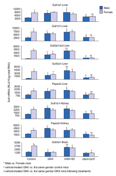
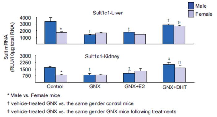
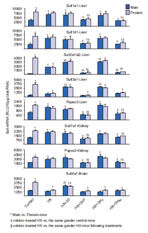
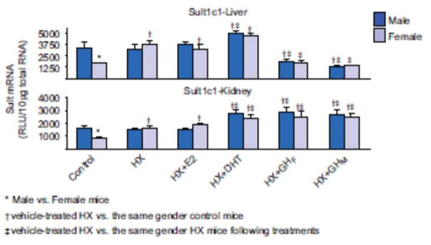
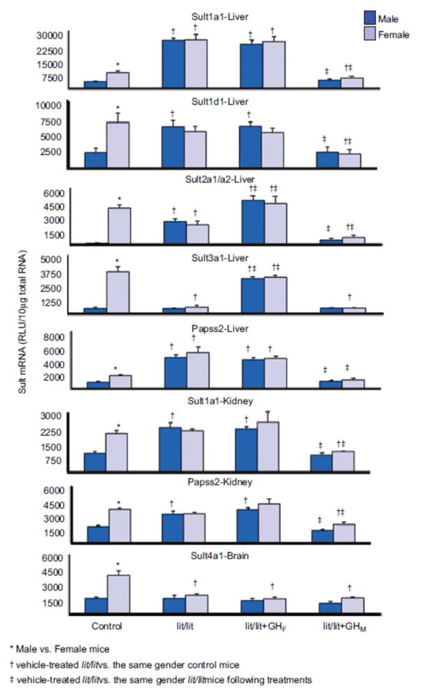
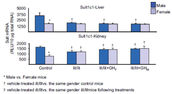
1. Marked gender differences in the expression of sulfotransferases (Sults) are known to exist in several species including rats, mice and hamsters. However, the mechanism for this gender difference is not known. Therefore, in the present study, it was determined whether sex and/or growth hormone (GH) are responsible for the gender difference in the expression of Sults using gonadectomized (GNX), hypophysectomized (HX) and GH-releasing hormone receptor-deficient little (lit/lit) mouse models. 2. Sult1a1 and Papss2 in liver and kidney, and Sult1d1 in liver are female-predominant in mice because of suppressive effects of both androgens and male-pattern GH secretion. Sult2a1/a2 is the most markedly female-predominant Sult in mouse liver due to suppressive effects of androgens and male-pattern GH secretion, as well as stimulatory effects by estrogens and female-pattern GH secretion. Sult3a1 is female-predominant in mouse liver due to suppressive effects of androgens as well as stimulatory effects of estrogens and female-pattern GH secretion. Sult1c1 expression is male-predominant in mouse liver and kidney because of stimulatory effects of androgens in males. Sult4a1 expression is female-predominant in mouse brain due to stimulatory effects of estrogens. 3. In conclusion, gender-divergent Sults are mostly female-predominant and Sult1c1 is the only male-dominant Sult. The gender differences in expression of various mouse Sults are influenced by various mechanisms involving sex and/or GHs.


Conflict of interest statement

DECLARATION OF INTEREST

This work was supported by NIH grants ES-09649, ES-09716, ES013714 and COBRE grant P20-RR-021940.

Figures

Figure 1
Figure 1
Effects of gonadectomy and sex-hormone replacements on the mRNA expression of female-predominant Sults in mice. Total liver, kidney or brain RNA were isolated and analyzed by bDNA for each Sult mRNA content. The data are presented as mean RLU ± SEM (n = 5). GNX (vehicle administered to gonadectomized mice), GNX + DHT (5α-dihydroxytestosterone administered to GNX mice), and GNX + E2 (17β-estradiol administered to GNX mice). Asterisk (*) represents statistically significant differences (P < 0.05) between male and female mice; single dagger (†) represents statistically significant differences (P < 0.05) between control mice and the same gender, vehicle-treated GNX mice; and double dagger (‡) represents statistically significant differences (P < 0.05) between vehicle-treated GNX mice and the same gender, GNX mice administered DHT or E2.
Figure 2
Figure 2
Effects of gonadectomy and sex-hormone replacements on the mRNA expression of male-predominant Sult1c1 in mice. Total liver or kidney RNA were isolated and analyzed by bDNA for each Sult mRNA content. The data Sare presented as mean RLU ± SEM (n = 5). GNX (vehicle administered to gonadectomized mice), GNX + DHT (5α-dihydroxytestosterone administered to GNX mice), and GNX + E2 (17β-estradiol administered to GNX mice). Asterisk (*) represents statistically significant differences (P < 0.05) between male and female mice; single dagger (†) represents statistically significant differences (P < 0.05) between control mice and the same gender, vehicle-treated GNX mice; and double dagger (‡) represents statistically significant differences (P < 0.05) between vehicle-treated GNX mice and the same gender, GNX mice administered DHT or E2.
Figure 3
Figure 3
Effects of hypophysectomy and hormone replacements on the mRNA expression of female-predominant Sults in mice. Total liver, kidney, or brain RNA were isolated and analyzed by bDNA for each Sult mRNA content. The data are presented as mean RLU ± SEM (n = 5). HX (placebo administered to hypophysectomized mice), HX + GHM (rat GH twice daily administered by i.p. injection to HX mice mimicking male-pattern GH secretion), HX + GHF (continuous infusion to HX mice via s.c. implanted 21-day-release 1-mg rat GH pellet mimicking female-pattern GH secretion), HX + DHT (5α-dihydroxytestosterone administered to HX mice), and HX + E2 (17β-estradiol administered to HX mice). Asterisk (*) represents statistically significant differences (P < 0.05) between male and female mice; single dagger (†) represents statistically significant differences (P < 0.05) between control mice and the same gender, vehicle-treated HX mice; and double dagger (‡) represents statistically significant differences (P < 0.05) between vehicle-treated HX mice and the same gender, HX mice following hormone replacement treatments.
Figure 4
Figure 4
Effects of hypophysectomy and hormone replacements on the mRNA expression of male-predominant Sult1c1 in mice. Total liver or kidney brain RNA were isolated and analyzed by bDNA for each Sult mRNA content. The data are presented as mean RLU ± SEM (n = 5). HX (placebo administered to hypophysectomized mice), HX + GHM (rat GH twice daily administered by i.p. injection to HX mice mimicking male-pattern GH secretion), HX + GHF (continuous infusion to HX mice via s.c. implanted 21-day-release 1-mg rat GH pellet mimicking female-pattern GH secretion), HX + DHT (5α-dihydroxytestosterone administered to HX mice), and HX + E2 (17β-estradiol administered to HX mice). Asterisk (*) represents statistically significant differences (P < 0.05) between male and female mice; single dagger (†) represents statistically significant differences (P < 0.05) between control mice and the same gender, vehicle-treated HX mice; and double dagger (‡) represents statistically significant differences (P < 0.05) between vehicle-treated HX mice and the same gender, HX mice following hormone replacement treatments.
Figure 5
Figure 5
Effects of GH secretion pattern on the mRNA expression of female-predominant Sults in mice. Total liver, kidney, or brain RNA were isolated and analyzed bybDNA for each Sult mRNA content. The data are presented as mean RLU ± SEM (n = 5). lit/lit, placebo administered to lit/lit mice; lit/lit + GHM, rat GH twice daily administered by i.p. injection to lit/lit mice mimicking male-pattern GH secretion; lit/lit + GHF, continuous infusion to lit/lit mice via s.c. implanted 21-day-release 1-mg rat GH pellet mimicking female-pattern GHsecretion. Asterisk (*) represents statistically significant differences (P < 0.05) between male and female mice; single dagger (†) represents statistically significant differences (P < 0.05) between control mice and the same gender, vehicle-treated lit/lit mice; and double dagger (‡) represents statistically significant differences (P < 0.05) between vehicle-treated lit/lit mice and the same gender lit/lit mice following GH replacement treatments.
Figure 6
Figure 6
Effects of GH secretion pattern on the mRNA expression of male-predominant Sult1c1 in mice. Total liver, or kidney RNA were isolated and analyzed by bDNA for each Sult mRNA content. The data are presented as mean RLU ± SEM (n = 5). lit/lit, placebo administered to lit/lit mice; lit/lit + GHM, rat GH twice daily administered by i.p. injection to lit/lit mice mimicking male-pattern GH secretion; lit/lit + GHF, continuous infusion to lit/lit mice via s.c. implanted 21-day-release 1-mg rat GH pellet mimicking female-pattern GH secretion. Asterisk (*) represents statistically significant differences (P < 0.05) between male and female mice; single dagger (†) represents statistically significant differences (P < 0.05) between control mice and the same gender, vehicle-treated lit/lit mice; and double dagger (‡) represents statistically significant differences (P < 0.05) between vehicle-treated lit/lit mice and the same gender lit/lit mice following GH replacement treatments.

Similar articles

Cited by

References

    1. Ahluwalia A, Clodfelter KH, Waxman DJ. Sexual dimorphism f rat liver gene expression: regulatory role of growth hormone revealed by deoxyribonucleic Acid microarray analysis. Mol Endocrinol. 2004;18:747–760. - PubMed
    1. Aksoy IA, Sochorová V, Weinshilboum RM. Human liver dehydroepiandrosterone sulfotransferase: nature and extent of individual variation. Clin Pharmacol Ther. 1993;54:498–506. - PubMed
    1. Alnouti Y, Klaassen CD. Tissue distribution and ontogeny of sulfotransferase enzymes in mice. Toxicol Sci. 2006;93:242–255. - PubMed
    1. Alnouti Y, Klaassen CD. Regulation of sulfotransferase enzymes by prototypical microsomal enzyme inducers in mice. J Pharmacol Exp Ther. 2008;324:612–621. - PubMed
    1. Balistreri WF, Zimmer L, Suchy FJ, Bove KE. Bile salt sulfotransferase: alterations during maturation and non-inducibility during substrate ingestion. J Lipid Res. 1984;25:228–235. - PubMed

Publication types

LinkOut - more resources