Review
. 2010 Dec;10(9):802-23.
doi: 10.2174/156652410793937813.

The retinal pigment epithelium in health and disease

Affiliations
Review

The retinal pigment epithelium in health and disease

J R Sparrow et al. Curr Mol Med. 2010 Dec.

Abstract

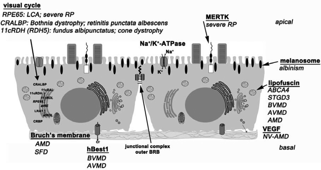
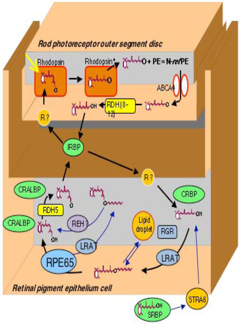
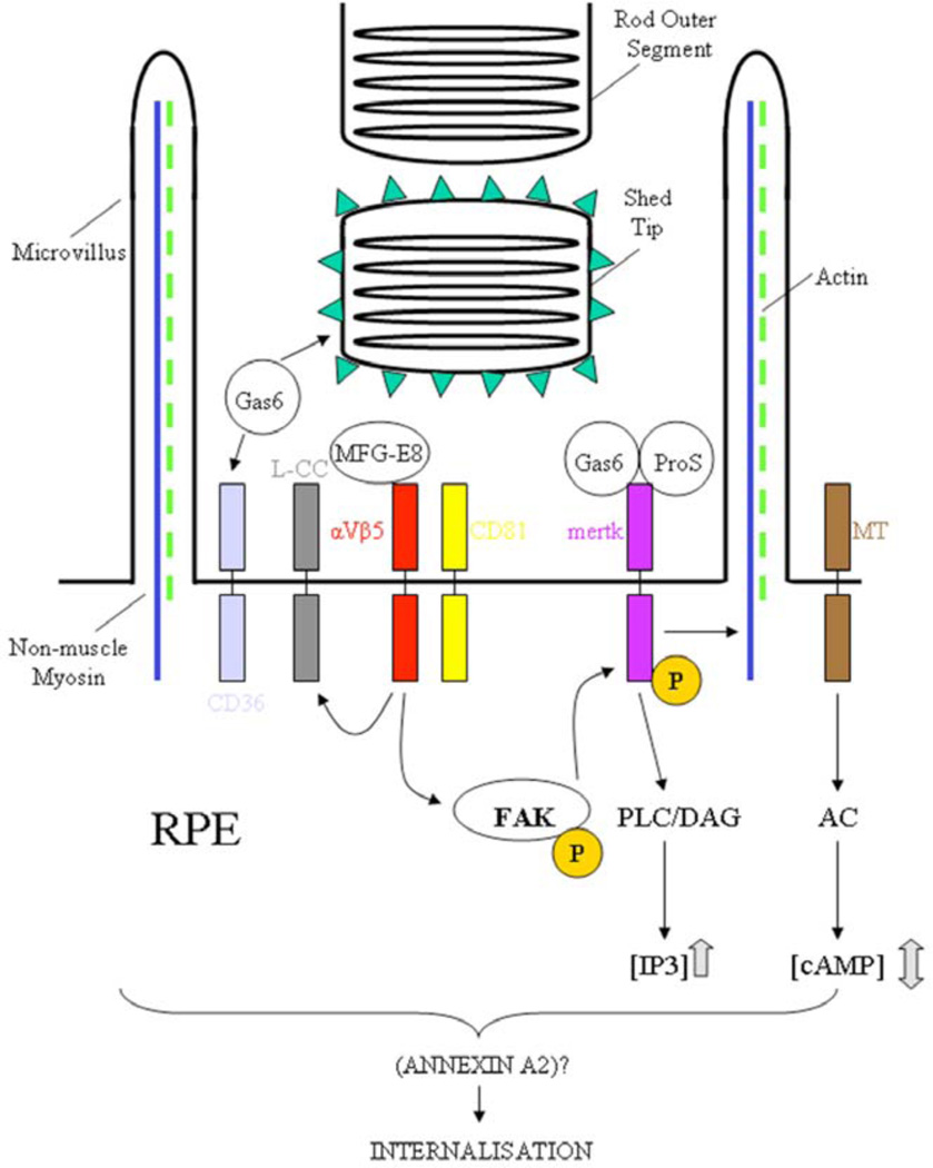
Retinal pigment epithelial cells (RPE) constitute a simple layer of cuboidal cells that are strategically situated behind the photoreceptor (PR) cells. The inconspicuousness of this monolayer contrasts sharply with its importance [1]. The relationship between the RPE and PR cells is crucial to sight; this is evident from basic and clinical studies demonstrating that primary dysfunctioning of the RPE can result in visual cell death and blindness. RPE cells carry out many functions including the conversion and storage of retinoid, the phagocytosis of shed PR outer segment membrane, the absorption of scattered light, ion and fluid transport and RPE-PR apposition. The magnitude of the demands imposed on this single layer of cells in order to execute these tasks, will become apparent to the reader of this review as will the number of clinical disorders that take origin from these cells.


Figures

Fig. (1)
Fig. (1). Schematic depiction of RPE cell structure / functions with associated retinal disorders
Cellular retinol binding protein (CRBP) binds all-trans-retinol (atROL) and directs it the smooth endoplasmic reticulum where lecithin:retinol acyltransferase (LRAT) converts all-trans-retinol to all-trans-retinyl ester which is then converted to 11-cis-retinol (11cROL) by RPE65 and to 11-cis retinal (11cRAL) by 11-cis-retinol dehydrogenase (11cRDH). Visual cycle proteins associated with forms of retinal degeneration include RPE65 (Leber congenital amaurosis, LCA; severe retinitis pigmentosa, RP); CRALBP and 11cRDH (encoded by gene retinol dehydrogenase 5, RDH5). The sodium/potassium adenosine triphosphatase (Na+/K+-ATPase) mediates outflux of sodium (Na+) and influx of potassium (K+). The c-mer tyrosine kinase (MERTK) receptor is responsible for internalization of shed out segment membrane. Mutations in MERTK are associated with severe retinitis pigmentosa (RP). Melanin pigment is contained within melanosomes; disruptions in melanin or melanosome biogenesis leads to albinism. Lipofuscin accumulates with excess in ABCA4-related disease, Stargardt-like macular dystrophy (STGD3), Best vitelliform macular dystrophy (BVMD) and adult-onset vitelliform macular dystrophy (AVMD); lipofuscin is also implicated in the pathogenesis of AMD. RPE cell secretion of vascular endothelial growth factor (VEGF) is implicated in neovascular age-related macular degeneration (NV-AMD). Junctional complexes between adjacent RPE cells constitute the outer blood-retinal barrier. Mutations in the protein bestrophin (hBEST1) are associated with Best vitelliform macular dystrophy (BVMD) and adult-onset vitelliform macular dystrophy (AVMD). The basal surface of the RPE rests on a basement membrane - Bruch’s membrane - that is implicated in age-related macular degeneration (AMD) and Sorby’s fundus dystrophy (SFD). See text for details.
Fig. (2)
Fig. (2). The visual cycle
Schematic of the visual cycle in rod photoreceptor outer segments and retinal pigment epithelium. Binding proteins are in green (interphotoreceptor retinol binding protein, IRBP; cellular retinol binding protein, CRBP; cellular retinaldehyde binding protein, CRALBP; serum retinol binding protein, SRBP); retinol dehydrogenases (RDH) in yellow (11-cis retinol dehydrogenase, RDH5); retinylester hydrolase (REH) in purple and other enzymes in blue (lecithin:retinol acyltransferase, LRAT; retinal G protein receptor, RGR; RPE65; membrane receptors (R) are brown (stimulated by retinoic acid gene 6, STRA6). N-retinylidene-phosphatidylethanolamine, N-ret-PE. See text for details.
Fig. (3)
Fig. (3). Fundi of patients with various conditions related to cycle visual genes
A, B: Left eye of a patient with Leber congenital amaurosis caused by mutations in RPE65 at 13 (A) and 22 (B) years of age. Note that fundus looks quite normal at 13 except for slight narrowing of retinal vessels and loss of the normal retinal reflex, while at 22 macular mottling (white arrow) and a few pigment deposits in periphery (black arrows) are present. C, D: Left eye of a 58 year-old female with fundus albipunctatus caused by mutations in RDH5. On fundus (C), note the presence of radially distributed, evenly shaped, dot-like white deposits that are characteristic of the disease, and macular pigment mottling (white arrow) which indicates macular involvement; on autofluorescence test (D), there is perifoveal hypofluorescence (white arrow) and the dot-like deposits which are not fluorescent appear as dark small spots (black arrow). E, F: Right eye of 13 year-old (E) and 24 year-old (F) males with retinitis punctata albescens caused by mutations in RLBP1. Note the presence of unevenly shaped and distributed, dot-like white deposits while there is normal macular reflex (white arrows).
Fig. (4)
Fig. (4). RPE phagocytosis
Schematic flow chart depicting the ligands (Gas6; protein S, ProS; milk fat globule-EGF 8, MFG-E8), receptors (c-mer tyrosine kinase, mertk; αVβ5; CD36; CD81; melatonin receptor, MT; and L-type calcium channels, L-CC) and intracellular messenger elements (adenylyl cyclase, AC; focal adhesion kinase, FAK; phospholipase C, PLC; diacylglycerol, DAG; inositol trisphosphate, IP3). P, phosphorylated sites; triangles on shed ROS surface represent phosphatidyl serine.

Similar articles

Cited by

References

    1. Bok D, Hall MO. The role of the pigment epithelium in the etiology of inherited retinal dystrophy in the rat. J Cell Biol. 1971;49:664–682. - PMC - PubMed
    1. Anderson DH, Fisher SK. The relationship of primate foveal cones to the pigment epithelium. J Ultrastruct Res. 1979;67:23–32. - PubMed
    1. Miller SS, Steinberg RH. Passive ionic properties of frog retinal pigment epithelium. J Membr Biol. 1977;36:337–372. - PubMed
    1. Shin K, Fogg VC, Margolis B. Tight junctions and cell polarity. Ann Rev Cell Develop Biol. 2006;22:207–235. - PubMed
    1. Rizzolo LJ. Development and role of tight junctions in the retinal pigment epithelium. Int Rev Cytol. 2007;258:195–234. - PubMed

MeSH terms