. 2011 Feb;103(2):55-68.
doi: 10.1042/BC20100111.

CDC25B associates with a centrin 2-containing complex and is involved in maintaining centrosome integrity

Affiliations

CDC25B associates with a centrin 2-containing complex and is involved in maintaining centrosome integrity

Rose Boutros et al. Biol Cell. 2011 Feb.

Abstract

Background information: CDC25 (cell division cycle 25) phosphatases function as activators of CDK (cyclin-dependent kinase)-cyclin complexes to regulate progression through the CDC. We have recently identified a pool of CDC25B at the centrosome of interphase cells that plays a role in regulating centrosome numbers.

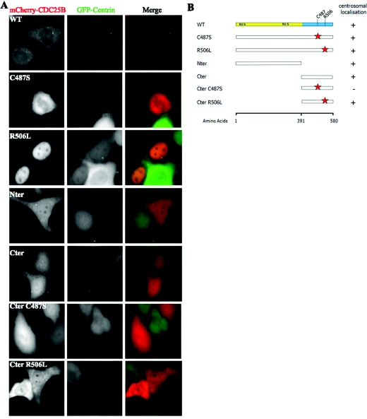
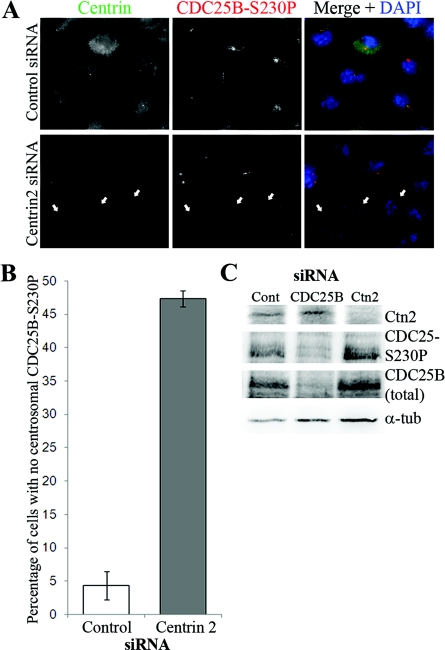
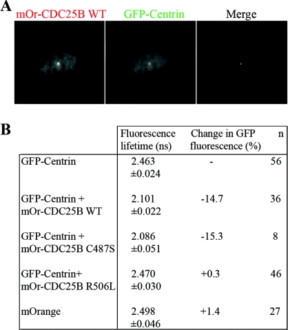
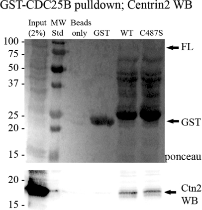
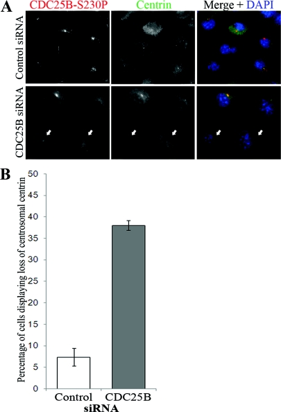
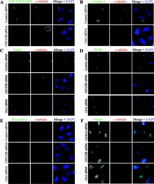
Results: In the present study, we demonstrate that CDC25B forms a close association with Ctn (centrin) proteins at the centrosome. This interaction involves both N- and C-terminal regions of CDC25B and requires CDC25B binding to its CDK-cyclin substrates. However, the interaction is not dependent on the enzyme activity of CDC25B. Although CDC25B appears to bind indirectly to Ctn2, this association is pertinent to CDC25B localization at the centrosome. We further demonstrate that CDC25B plays a role in maintaining the overall integrity of the centrosome, by regulating the centrosome levels of multiple centrosome proteins, including that of Ctn2.

Conclusions: Our results therefore suggest that CDC25B associates with a Ctn2-containing multiprotein complex in the cytoplasm, which targets it to the centrosome, where it plays a role in maintaining the centrosome levels of Ctn2 and a number of other centrosome components.


Figures

Figure 1
Figure 1. Both N- and C-terminal domains of CDC25B are involved in its centrosome targeting
Various mutant forms of mCherry-CDC25B were derived, transfected into HeLa-Ctn1 cells and analysed for their ability to localize to the centrosome. (A) Examples of HeLa-Ctn1 cell transfected with mCherry-WT, -R506L, -Nter or -Cter CDC25B constructs. (B) Schematic diagram representing various N- (regulatory, yellow) and C- (catalytic, blue) domain mutant forms of CDC25B, in addition to mutations of specific amino acids within the catalytic domain (red stars), representing the mutation of Cys487 [critical for CDC25B catalytic activity (Gabrielli et al., 1996)] to serine (C487S), or Arg506 [critical for substrate binding (Sohn et al., 2004)] to leucine (R506L). The right-hand column indicates the presence (+) or absence (−) of each mutant at the centrosome 24 h post-transfection in HeLa-Ctn1 cells.
Figure 2
Figure 2. Ctn2 depletion causes loss of CDC25B from the centrosome
HeLa cells were treated with scrambled control or Ctn2 siRNA duplexes for 48 h before immunofluorescence detection of Ctn (green) and CDC25B (CDC25B-S230P, red). (A) Examples of Ctn2-depleted cells also demonstrating a detectable loss of centrosomal CDC25B (arrows) in comparison with cells treated with control siRNA duplexes. (B) Percentage of HeLa cells in which CDC25B-S230P and Ctn2 (Ctn2 siRNA only) were not detectable or substantially diminished from the centrosomes. In Ctn2 siRNA-treated cells, only the cells displaying a loss of centrosomal Ctn2 were scored for the presence or significant loss of centrosomal CDC25B-S230P signal. Bars represent mean results for at least 200 cells counted from three independent experiments ±S.D. (C) Western blots of lysates from control (Cont), Ctn2 and CDC25B siRNA-depleted cells immunoblotted for Ctn2, total CDC25B (using the C20 antibody from Santa Cruz), CDC25B-S230P and α-tubulin (α-tub) as loading control.
Figure 3
Figure 3. WT CDC25B interacts with Ctn1 at the centrosome in live cells
FRET–FLIM analyses of HeLa-Ctn1 cells co-expressing mOrange-CDC25B. (A) Example of a HeLa-Ctn1 cell co-transfected with mOrange (mOr)-CDC25B WT, demonstrating co-localization with GFP-Ctn1. (B) GFP fluorescent lifetime values in HeLa-Ctn1 cells that were either mock-transfected (GFP-Ctn) or co-transfected with mOrange vector control, WT CDC25B (mOr-CDC25B WT), the catalytically inactive mutant mOr-CDC25B C487S, or the substrate-binding mutant mOr-CDC25B R506L. Fluorescence lifetime measurements are in nanoseconds (ns) and n represents the total number of cells analysed from three to five independent experiments.
Figure 4
Figure 4. Endogenous Ctn2 can be pulled down with GST–CDC25B
GST–CDC25B fusion proteins produced in bacteria were bound to GST beads and incubated with protein extracts from HeLa cells. Proteins attached to the GST beads were denatured and separated by SDS/PAGE. (Upper panel) Ponceau stain of nitrocellulose showing levels of GST or the various GST–CDC25B fusion proteins in each reaction. (Lower panel) Western-blot analysis of the same nitrocellulose membrane, immunostained for Ctn2.
Figure 5
Figure 5. CDC25B and Ctn2 centrosomal localizations are interdependent
HeLa cells were treated with scrambled control or CDC25B siRNA duplexes for 48 h before immunofluorescence detection of CDC25B (CDC25B-S230P) and Ctn. (A) Examples of siRNA-treated cells from which CDC25B was successfully depleted which also show a loss of centrosomal Ctn2 (arrows). (B) Percentage of HeLa cells displaying loss of both CDC25B-S230P (CDC25B siRNA only) and Ctn2 from the centrosomes. Bars represent means for at least 200 cells counted from three independent experiments ±S.D.
Figure 6
Figure 6. CDC25B depletion causes a loss of centrosome integrity in HeLa cells
HeLa cells were treated with scrambled control, CDC25B or Ctn2 siRNA duplexes, and co-stained for γ-tubulin (red) and 4′, 6-diamino-2-phenylindole (DAPI, blue), along with (A) CDC25B (CDC25B-S230P), (B) Ctn2, (C) Nedd1, (D) PCM1, (E) pericentrin or (F) ninein (green).

References

    1. Araki M., Masutani C., Takemura M., Uchida A., Sugasawa K., Kondoh J., Ohkuma Y., Hanaoka F. Centrosome protein centrin 2/caltractin 1 is part of the xeroderma pigmentosum group C complex that initiates global genome nucleotide excision repair. J. Biol. Chem. 2001;276:18665–18672. - PubMed
    1. Azimzadeh J., Bornens M. Structure and duplication of the centrosome. J. Cell Sci. 2007;120:2139–2142. - PubMed
    1. Azimzadeh J., Hergert P., Delouvee A., Euteneuer U., Formstecher E., Khodjakov A., Bornens M. hPOC5 is a centrin-binding protein required for assembly of full-length centrioles. J. Cell Biol. 2009;185:101–114. - PMC - PubMed
    1. Baron A.T., Suman V.J., Nemeth E., Salisbury J.L. The pericentriolar lattice of PtK2 cells exhibits temperature and calcium-modulated behavior. J. Cell Sci. 1994;107:2993–3003. - PubMed
    1. Baum P., Furlong C., Byers B. Yeast gene required for spindle pole body duplication: homology of its product with Ca2+-binding proteins. Proc. Natl. Acad. Sci. U.S.A. 1986;83:5512–5516. - PMC - PubMed

Publication types