

Distinct glycan-charged phosphodolichol carriers are required for the assembly of the pentasaccharide N-linked to the Haloferax volcanii S-layer glycoprotein
- PMID: 21091511
- PMCID: PMC3074503
- DOI: 10.1111/j.1365-2958.2010.07405.x
Distinct glycan-charged phosphodolichol carriers are required for the assembly of the pentasaccharide N-linked to the Haloferax volcanii S-layer glycoprotein
Abstract
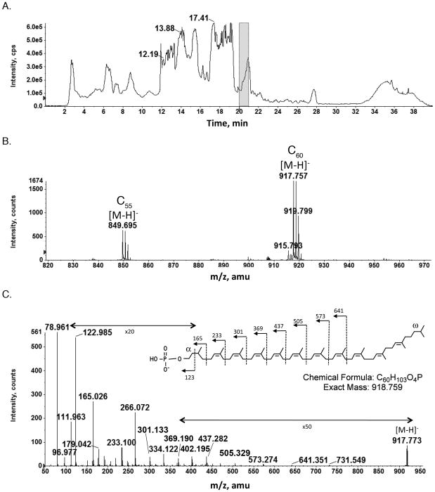
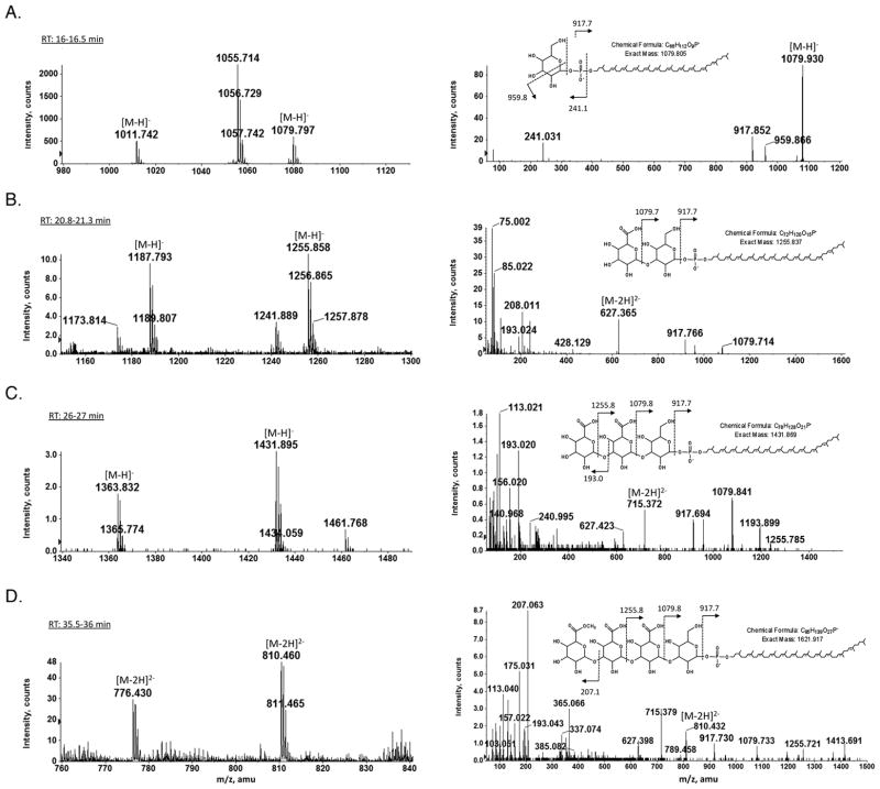
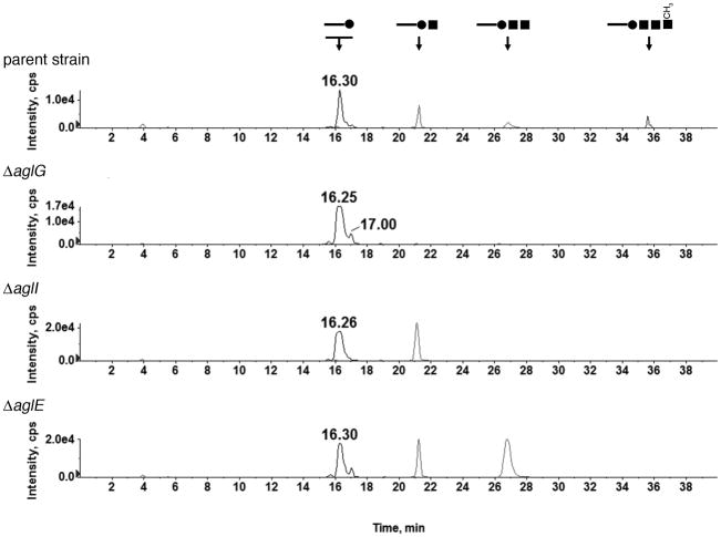
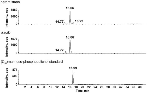
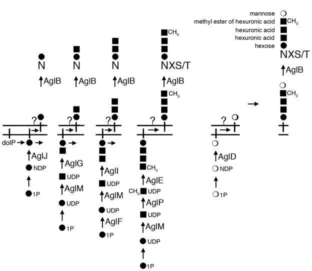
In Archaea, dolichol phosphates have been implicated as glycan carriers in the N-glycosylation pathway, much like their eukaryal counterparts. To clarify this relation, highly sensitive liquid chromatography/mass spectrometry was employed to detect and characterize glycan-charged phosphodolichols in the haloarchaeon Haloferax volcanii. It is reported that Hfx. volcanii contains a series of C(55) and C(60) dolichol phosphates presenting saturated isoprene subunits at the α and ω positions and sequentially modified with the first, second, third and methylated fourth sugar subunits comprising the first four subunits of the pentasaccharide N-linked to the S-layer glycoprotein, a reporter of N-glycosylation. Moreover, when this glycan-charged phosphodolichol pool was examined in cells deleted of agl genes encoding glycosyltransferases participating in N-glycosylation and previously assigned roles in adding pentasaccharide residues one to four, the composition of the lipid-linked glycans was perturbed in the identical manner as was S-layer glycoprotein N-glycosylation in these mutants. In contrast, the fifth sugar of the pentasaccharide, identified as mannose in this study, is added to a distinct dolichol phosphate carrier. This represents the first evidence that in Archaea, as in Eukarya, the oligosaccharides N-linked to glycoproteins are sequentially assembled from glycans originating from distinct phosphodolichol carriers.
© 2010 Blackwell Publishing Ltd.
Figures
Similar articles
-
Assembling Glycan-Charged Dolichol Phosphates: Chemoenzymatic Synthesis of a Haloferax volcanii N-Glycosylation Pathway Intermediate.Bioconjug Chem. 2017 Sep 20;28(9):2461-2470. doi: 10.1021/acs.bioconjchem.7b00436. Epub 2017 Aug 30. Bioconjug Chem. 2017. PMID: 28809486
-
AglR is required for addition of the final mannose residue of the N-linked glycan decorating the Haloferax volcanii S-layer glycoprotein.Biochim Biophys Acta. 2012 Oct;1820(10):1664-70. doi: 10.1016/j.bbagen.2012.06.014. Epub 2012 Jun 27. Biochim Biophys Acta. 2012. PMID: 22750201 Free PMC article.
-
AglJ adds the first sugar of the N-linked pentasaccharide decorating the Haloferax volcanii S-layer glycoprotein.J Bacteriol. 2010 Nov;192(21):5572-9. doi: 10.1128/JB.00705-10. Epub 2010 Aug 27. J Bacteriol. 2010. PMID: 20802039 Free PMC article.
-
Lipid sugar carriers at the extremes: The phosphodolichols Archaea use in N-glycosylation.Biochim Biophys Acta Mol Cell Biol Lipids. 2017 Jun;1862(6):589-599. doi: 10.1016/j.bbalip.2017.03.005. Epub 2017 Mar 19. Biochim Biophys Acta Mol Cell Biol Lipids. 2017. PMID: 28330764 Free PMC article. Review.
-
Add salt, add sugar: N-glycosylation in Haloferax volcanii.Biochem Soc Trans. 2013 Feb 1;41(1):432-5. doi: 10.1042/BST20120142. Biochem Soc Trans. 2013. PMID: 23356324 Review.
Cited by
-
Influence of N-Glycosylation on Virus-Host Interactions in Halorubrum lacusprofundi.Viruses. 2023 Jun 28;15(7):1469. doi: 10.3390/v15071469. Viruses. 2023. PMID: 37515157 Free PMC article.
-
Crystal structures of an archaeal oligosaccharyltransferase provide insights into the catalytic cycle of N-linked protein glycosylation.Proc Natl Acad Sci U S A. 2013 Oct 29;110(44):17868-73. doi: 10.1073/pnas.1309777110. Epub 2013 Oct 14. Proc Natl Acad Sci U S A. 2013. PMID: 24127570 Free PMC article.
-
AglS, a novel component of the Haloferax volcanii N-glycosylation pathway, is a dolichol phosphate-mannose mannosyltransferase.J Bacteriol. 2012 Dec;194(24):6909-16. doi: 10.1128/JB.01716-12. Epub 2012 Oct 19. J Bacteriol. 2012. PMID: 23086206 Free PMC article.
-
Identification of Haloferax volcanii Pilin N-Glycans with Diverse Roles in Pilus Biosynthesis, Adhesion, and Microcolony Formation.J Biol Chem. 2016 May 13;291(20):10602-14. doi: 10.1074/jbc.M115.693556. Epub 2016 Mar 10. J Biol Chem. 2016. PMID: 26966177 Free PMC article.
-
N-Glycosylation Is Important for Proper Haloferax volcanii S-Layer Stability and Function.Appl Environ Microbiol. 2017 Mar 2;83(6):e03152-16. doi: 10.1128/AEM.03152-16. Print 2017 Mar 15. Appl Environ Microbiol. 2017. PMID: 28039139 Free PMC article.
References
-
- Abu-Qarn M, Eichler J. Protein N-glycosylation in Archaea: defining Haloferax volcanii genes involved in S-layer glycoprotein glycosylation. Mol Microbiol. 2006;61:511–525. - PubMed
-
- Abu-Qarn M, Yurist-Doutsch S, Giordano A, Trauner A, Morris HR, Hitchen P, et al. Haloferax volcanii AglB and AglD are involved in N-glycosylation of the S-layer glycoprotein and proper assembly of the surface layer. J Mol Biol. 2007;14:1224–1236. - PubMed
-
- Burda P, Aebi M. The dolichol pathway of N-linked glycosylation. Biochim Biophys Acta. 1999;1426:239–257. - PubMed
-
- Calo D, Kaminski L, Eichler J. Protein glycosylation in Archaea: Sweet and extreme. Glycobiology. 2010;20:1065–1079. - PubMed
Publication types
MeSH terms
Substances
Grants and funding
LinkOut - more resources
Full Text Sources
Molecular Biology Databases
