. 2011 Nov;15(11):2343-52.
doi: 10.1111/j.1582-4934.2010.01215.x.

Immunosuppressive effect of bone marrow-derived mesenchymal stem cells in inflammatory microenvironment favours the growth of B16 melanoma cells

Affiliations

Immunosuppressive effect of bone marrow-derived mesenchymal stem cells in inflammatory microenvironment favours the growth of B16 melanoma cells

Zhipeng Han et al. J Cell Mol Med. 2011 Nov.

Abstract

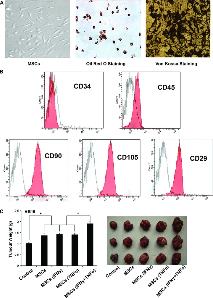
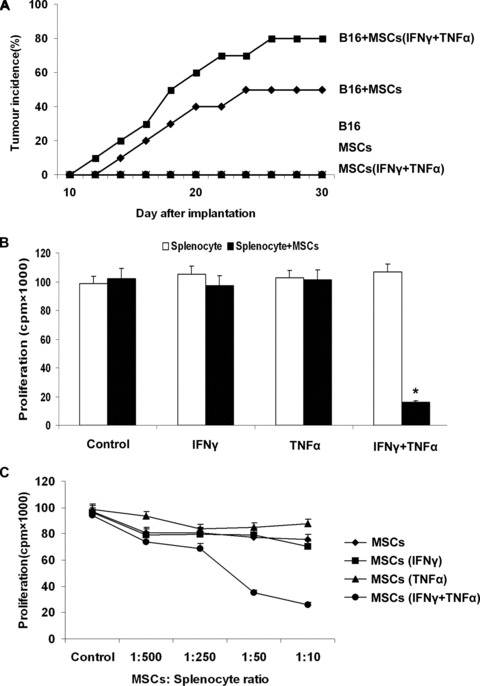
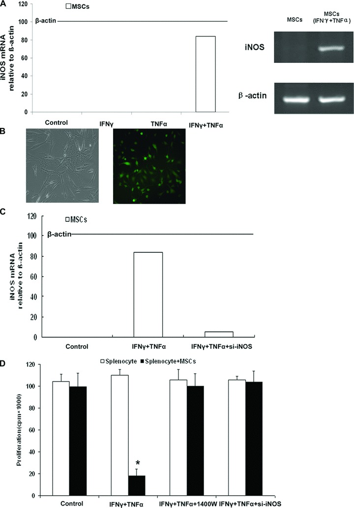
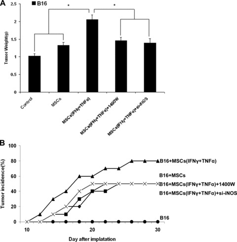
Mesenchymal stem cells (MSCs) are studied for their potential clinical use in regenerative medicine, tissue engineering and tumour therapy. However, the therapeutic application of MSCs in tumour therapy still remains limited unless the immunosuppressive role of MSCs for tumour growth in vivo is better understood. In this study, we investigated the mechanism of MSCs favouring tumour escape from immunologic surveillance in inflammatory microenvironment. We first compared the promotive capacity of bone marrow-derived MSCs on B16 melanoma cells growth in vivo, pre-incubated or not with the inflammatory cytokines interferon (IFN)-γ and tumour necrosis factor (TNF)-α. We showed that the development of B16 melanoma cells is faster when co-injected with MSCs pre-incubated with IFN-γ and TNF-α compared with control groups. Moreover, tumour incidence increases obviously in allogeneic recipients when B16 melanoma cells were co-injected with MSCs pre-incubated with IFN-γ and TNF-α. We then demonstrated that the immunosuppressive function of MSCs was elicited by IFN-γ and TNF-α. These cytokine combinations provoke the expression of inducible nitric oxide synthase (iNOS) by MSCs. The impulsive effect of MSCs treated with inflammatory cytokines on B16 melanoma cells in vivo can be reversed by inhibitor or short interfering RNA of iNOS. Our results suggest that the MSCs in tumour inflammatory microenvironment may be elicited of immunosuppressive function, which will help tumour to escape from the immunity surveillance.


Figures

Fig 1
Fig 1
MSCs treated with proinflammatory cytokines favour the growth of tumour. (A) MSCs at passage 8 were grown under different conditions that favour differentiation into either adipocytes (for 14 days), or osteoblasts (for 14 days), as described in ‘Materials and methods’. The presence of triglycerides, characteristic of adipocytes, was revealed by staining with oil red O. Calcium deposition, indicative of osteoblasts, was stained with Von Kossa stain. (B) MSCs derived from C57BL/6 mice were stained with commercially available antibodies to analysis the surface marker by flow cytometry (red). Corresponding antibodies of the same isotype were used as controls (white). (C) C57BL/6 MSCs (1 × 10⁶) were pretreated with proinflammatory cytokines IFN-γ and TNF-α (20 ng/ml each) for 12 hrs and then mixed with B16 melanoma cells (5 × 10⁶) to perform subcutaneous administration in the C57BL/6 mice armpit area. After 14 days of implantation, the animals were sacrificed and tumours were dissected. The weight and volume of B16 tumours were measured. The weight of tumour were measured after been removed from the mice. (*P < 0.05).
Fig 2
Fig 2
The rejection from Balb/c mice on B16 melanoma cells are reduced by the MSCs treated with proinflammatory cytokines. (A) Balb/c MSCs (1 × 10⁶) were pretreated with inflammatory cytokines IFN-γ and TNF-α (20 ng/ml each) for 12 hrs and then mixed with B16 melanoma cells (5 × 10⁶) to perform subcutaneous administration in the Balb/c mice armpit area. Tumour incidence was observed to evaluate the immunosuppressive function of MSCs that assisted the B16 melanoma cells from escaping the immunological rejection of Balb/c mice. As negative controls, B16 cells or MSCs alone were injected in Balb/c mice. (B) C57BL/6 MSCs were pretreated with inflammatory cytokines IFN-γ and TNF-α (20 ng/ml each) for 12 hrs. Fresh C57BL/6 splenocytes were activated by ConA (5 μg/ml) for 72 hrs and IL-2 (20 ng/ml) was added for proliferation. MSCs were cocultured in a 96-well with splenocytes (1 × 10⁵/well) plate at a 1:10 ratio and cell proliferation was assessed 72 hrs later by 3H-Tdr incorporation. (C) MSCs were mixed with splenocytes (1 × 10⁵/well) at different ratios (1:500, 1:250, 1:50, 1:10), and the proliferation was assessed 72 hrs later (*P < 0.05).
Fig 3
Fig 3
Immunosuppression by MSCs requires nitric oxide. (A) C57BL/6 MSCs were pretreated with proinflammatory cytokines IFN-γ and TNF-α (20 ng/ml each) for 12 hrs. iNOS mRNA in MSCs was assayed by real-time PCR and compared to β-actin mRNA, defined as 100 arbitrary unit. The electropherogram of iNOS cDNA demonstrated the same result as real-time PCR. (B) Transfections of iNOS siRNA into MSCs were performed with a Lipofectamine, and FAM was observed under fluorescence microscope (right, ×200). (C) Real-time-PCR result showed that the expression of iNOS was markedly decreased in MSCssi-iNOS stimulated by both IFN-γ and TNF-α, the inhibitory efficiency was more than 90% compared with the MSCsvector stimulated by both IFN-γ and TNF-α. (D) MSCs from C57BL/6 mice were cocultured with fresh C57BL/6 splenocytes plus Con A and IL-2, with or without the iNOS inhibitor – 1400W (100 μM) and siRNA of iNOS. Cell proliferation was assayed by 3H-Tdr incorporation after 72 hrs.
Fig 4
Fig 4
The growth of tumour favoured by MSCs in vivo can be inhibited by iNOS inhibitor. (A) C57BL/6 MSCs (1 × 10⁶) were pretreated with inflammatory cytokines IFN-γ and TNF-α (20 ng/ml each) for 12 hrs and then mixed with B16 cells (5 × 10⁶) to perform subcutaneous administration in the C57BL/6 mice armpit area. On days 2, 5, 8 and 11 after implantation, recipients were injected i.p. with iNOS inhibitor – 1400W. After 14 days of implantation, the animals were sacrificed and tumours were dissected. The weight and volume of B16 tumours were measured. Transfections of iNOS siRNA into C57BL/6 MSCs were performed with a Lipofectamine. Then the MSCssi-iNOS stimulated by both IFN-γ and TNF-α for 12 hrs before been mix with B16 melanoma cells (5 × 10⁶) to perform subcutaneous administration in the C57BL/6 mice armpit area. After 14 days of implantation, the animals were killed and tumours were dissected. The weight and volume of B16 tumours were measured. (B) Balb/c MSCs (1 × 10⁶) were pretreated with inflammatory cytokines IFN-γ and TNF-α (20 ng/ml each) for 12 hrs and then mixed with B16 melanoma cells (5 × 10⁶) to perform subcutaneous administration in the Balb/c mice armpit area. On days 2, 5, 8 and 11 after implantation, recipients were injected i.p. with iNOS inhibitor – 1400W. Tumour incidence was observed. Transfections of iNOS siRNA into Balb/c MSCs were performed with a Lipofectamine. Then the MSCssi-iNOS stimulated by both IFN-γ and TNF-α for 12 hrs before been mixed with B16 melanoma cells (5 × 10⁶) to perform subcutaneous administration in the Balb/c mice armpit area. Tumour incidence was observed.

Similar articles

Cited by

References

    1. Pittenger MF, Mackay AM, Beck SC, et al. Multilineage potential of adult human mesenchymal stem cells. Science. 1999;284:143–7. - PubMed
    1. Barry FP, Murphy JM. Mesenchymal stem cells: clinical applications and biological characterization. Int J Biochem Cell Biol. 2004;36:568–84. - PubMed
    1. da Silva Meirelles L, Chagastelles PC, et al. Mesenchymal stem cells reside in virtually all post-natal organs and tissues. J Cell Sci. 2006;119:2204–13. - PubMed
    1. Noel D, Djouad F, Jorgense C. Regenerative medicine through mesenchymal stem cells for bone and cartilage repair. Curr Opin Investig Drugs. 2002;3:1000–4. - PubMed
    1. Sato K, Ozaki K, Oh I, et al. Nitric oxide plays a critical role in suppression of T-cell proliferation by mesenchymal stem cells. Blood. 2007;109:228–34. - PubMed

Publication types

MeSH terms