. 2011 Mar;162(5):1171-8.
doi: 10.1111/j.1476-5381.2010.01119.x.

Hydrogen sulphide protects mouse pancreatic β-cells from cell death induced by oxidative stress, but not by endoplasmic reticulum stress

Affiliations

Hydrogen sulphide protects mouse pancreatic β-cells from cell death induced by oxidative stress, but not by endoplasmic reticulum stress

S Taniguchi et al. Br J Pharmacol. 2011 Mar.

Abstract

Background and purpose: Hydrogen sulphide (H₂S), a potentially toxic gas, is also involved in the neuroprotection, neuromodulation, cardioprotection, vasodilatation and the regulation of inflammatory response and insulin secretion. We have recently reported that H₂S suppresses pancreatic β-cell apoptosis induced by long-term exposure to high glucose. Here we examined the protective effects of sodium hydrosulphide (NaHS), an H₂S donor, on various types of β-cell damage.

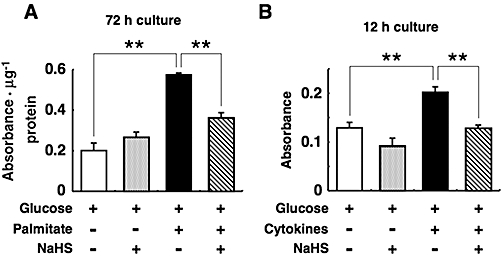
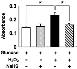
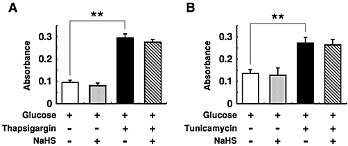
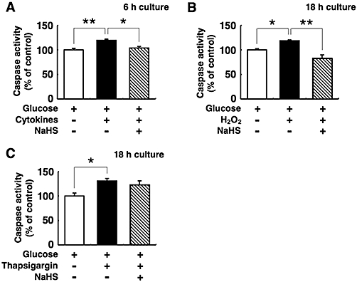
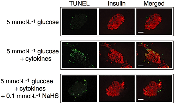
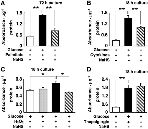
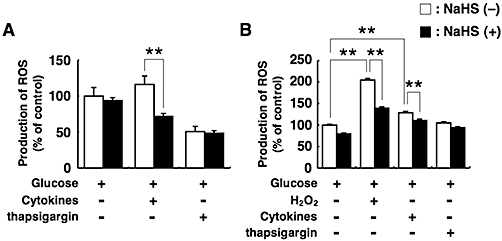
Experimental approach: Isolated islets from mice or the mouse insulinoma MIN6 cells were cultured with palmitate, cytokines (a mixture of tumour necrosis factor-α, interferon-γ and interleukin-1β), hydrogen peroxide, thapsigargin or tunicamycin with or without NaHS. We examined DNA fragmentation, caspase-3 and -7 activities and reactive oxygen species (ROS) production in the treated cells thereafter. Apoptotic cell death in isolated islets was also assessed by the terminal deoxynucleotidyl transferase-mediated deoxyuridine triphosphate nick end labelling (TUNEL) method.

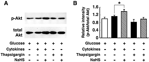
Key results: NaHS suppressed DNA fragmentation and the activities of caspase-3 and -7 induced by palmitate, the cytokines or hydrogen peroxide. In contrast, NaHS failed to protect islets and MIN6 cells from apoptosis induced by thapsigargin and tunicamycin, both of which cause endoplasmic reticulum stress. NaHS suppressed ROS production induced by cytokines or hydrogen peroxide but it had no effect on ROS production in thapsigargin-treated cells. NaHS increased Akt phosphorylation in MIN6 cells treated with cytokines but not in cells treated with thapsigargin. Treatment with NaHS decreased TUNEL-positive cells in cytokine-exposed islets.

Conclusions and implications: H₂S may prevent pancreatic β-cells from cell apoptosis via an anti-oxidative mechanism and the activation of Akt signalling.


Figures

Figure 1
Figure 1
Cytoprotective effects of sodium hydrosulphide (NaHS) against pancreatic islet cell death induced by palmitate (A) and cytokines (B). Mouse pancreatic islets were cultured for the indicated durations with 5 mmol·L−1 glucose alone or 5 mmol·L−1 glucose plus 0.5 mmol·L−1 palmitate or the cytokine mixture (1000 U·mL−1 tumour necrosis factor-α, 1000 U·mL−1 interferon-γ and 50 U·mL−1 interleukin-1β) in the presence or absence of 0.1 mmol·L−1 NaHS. After this, histone-associated DNA fragments were quantified by elisa to evaluate apoptotic cell death. Each column represents the mean ± SE from three to five separate experiments. **P < 0.01.
Figure 2
Figure 2
Protection by sodium hydrosulphide (NaHS) of pancreatic islets from oxidative stress induced by hydrogen peroxide (H2O2). Mouse pancreatic islets were cultured for the indicated durations with 5 mmol·L−1 glucose alone or 5 mmol·L−1 glucose plus 10 µmol·L−1 H2O2 in the presence or absence of 0.1 mmol·L−1 NaHS. After this, histone-associated DNA fragments were quantified by elisa to evaluate apoptotic cell death. Each column represents the mean ± SE from three to five separate experiments. *P < 0.05.
Figure 3
Figure 3
Lack of effect of sodium hydrosulphide (NaHS) on cell death induced by thapsigargin (A) or tunicamycin (B). Pancreatic islets were cultured for 18 h with or without 1 µmol·L−1 thapsigargin or 5 µg·mL−1 tunicamycin in the presence or absence of 0.1 mmol·L−1 NaHS. These experiments were carried out consistently in the presence of 5 mmol·L−1 glucose. After the culture, fragmented DNA was measured by elisa. Each column represents the mean ± SE from three to five separate experiments. *P < 0.05 and **P < 0.01.
Figure 4
Figure 4
Effects of sodium hydrosulphide (NaHS) on caspase-3 and -7 activities in pancreatic islets. Mouse pancreatic islets were cultured with 5 mmol·L−1 glucose alone or 5 mmol·L−1 glucose plus the cytokine mixture (A), 10 µmol·L−1 hydrogen peroxide (H2O2) (B) or 1 µmol·L−1 thapsigargin (C) with or without 0.1 mmol·L−1 NaHS. After the culturing, the activities of caspase-3 and -7 were analysed. Each column represents the mean ± SE from three to five separate experiments. *P < 0.05 and **P < 0.01.
Figure 5
Figure 5
Terminal deoxynucleotidyl transferase-mediated deoxyuridine triphosphate nick end labelling (TUNEL) staining of mouse islets. Isolated pancreatic islets were cultured for 18 h with 5 mmol·L−1 glucose alone (upper lane) or 5 mmol·L−1 glucose plus the cytokine mixture with (lower lane) or without (middle lane) 0.1 mmol·L−1 NaHS. Apoptotic cell death is demonstrated by the TUNEL staining in green. Insulin-positive cells are shown in red. Scale bar = 50 µm.
Figure 6
Figure 6
Effects of sodium hydrosulphide (NaHS) on MIN6 cell death induced by palmitate, cytokines, hydrogen peroxide (H2O2) or thapsigargin. MIN6 were cultured with 20 mmol·L−1 glucose alone or 20 mmol·L−1 glucose plus 0.5 mmol·L−1 palmitate (A), the cytokine mixture (B), 40 µmol·L−1 H2O2 (C) or 1 µmol·L−1 thapsigargin (D) in the presence or absence of 0.1 mmol·L−1 NaHS. Fragmented DNA in MIN6 cells was quantified by elisa to evaluate apoptotic cell death. Each column represents the mean ± SE from three to five separate experiments. *P < 0.05 and **P < 0.01.
Figure 7
Figure 7
Production of reactive oxygen species (ROS) in pancreatic islets and MIN6 cells. (A) Pancreatic islets were preincubated for 30 min with 10 µmol·L−1 dichlorodihydrofluorescein diacetate (H2DCFDA) fluorescein, and then incubated for 12 h with 5 mmol·L−1 glucose alone or 5 mmol·L−1 glucose plus the cytokine mixture or 1 µmol·L−1 thapsigargin in the presence or absence of 0.1 mmol·L−1 sodium hydrosulphide (NaHS). (B) MIN6 cells were preincubated for 30 min with 10 µmol·L−1 H2DCFDA fluorescein, and then incubated for 12 h with 20 mmol·L−1 glucose alone or 20 mmol·L−1 glucose plus 30 µmol·L−1 hydrogen peroxide (H2O2), the cytokine mixture or 1 µmol·L−1 thapsigargin in the presence or absence of 0.1 mmol·L−1 NaHS. The ROS levels were measured using a dichlorofluorescein fluorescence assay. Each column represents the mean ± SE from three to five separate experiments. **P < 0.01.
Figure 8
Figure 8
Effects of sodium hydrosulphide (NaHS) on Akt phosphorylation. MIN6 cells were cultured for 6 h with the cytokine mixture or 1 µmol·L−1 thapsigargin in the presence or absence of 0.1 mmol·L−1 NaHS. The concomitant concentration of glucose was 20 mmol·L−1. Protein extracts from these cells were separated by sodium dodecyl sulphate-polyacrylamide gel electrophoresis and immunoblotted for the detection of phosphorylated (upper lane) and total (lower lane) Akt (A). The phosphorylated Akt (p-Akt) and total Akt levels were then analysed by densitometrical measurement of the intensity of each band with the NIH image software (B). The ratio of p-Akt to total Akt was calculated. Each column represents the mean ± SE from four to five separate experiments. *P < 0.05.

References

    1. Assmann A, Ueki K, Winnay JN, Kadowaki T, Kulkarni RN. Glucose effects on beta-cell growth and survival require activation of insulin receptors and insulin receptor substrate 2. Mol Cell Biol. 2009;29:3219–3228. - PMC - PubMed
    1. Baskar R, Li L, Moore PK. Hydrogen sulfide-induces DNA damage and changes in apoptotic gene expression in human lung fibroblast cells. FASEB J. 2007;21:247–255. - PubMed
    1. Calvert JW, Jha S, Gundewar S, Elrod JW, Ramachandran A, Pattillo CB, et al. Hydrogen sulfide mediates cardioprotection through Nrf2 signaling. Circ Res. 2009;105:365–374. - PMC - PubMed
    1. Cao Y, Adhikari S, Ang AD, Bhatia M. Mechanism of induction of pancreatic acinar cell apoptosis by hydrogen sulfide. Am J Physiol Cell Physiol. 2006;291:C503–C510. - PubMed
    1. Chang-Chen KJ, Mullur R, Bernal-Mizrachi E. β-cell failure as a complication of diabetes. Rev Endocr Metab Disord. 2008;9:329–343. - PMC - PubMed

Publication types

MeSH terms