. 2011 Mar;162(6):1314-25.
doi: 10.1111/j.1476-5381.2010.01121.x.

Functional antagonism of β-adrenoceptor subtypes in the catecholamine-induced automatism in rat myocardium

Affiliations

Functional antagonism of β-adrenoceptor subtypes in the catecholamine-induced automatism in rat myocardium

D C Boer et al. Br J Pharmacol. 2011 Mar.

Abstract

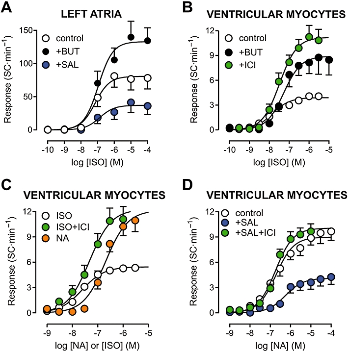
Background and purpose: Myocardial automatism and arrhythmias may ensue during strong sympathetic stimulation. We sought to investigate the relevant types of adrenoceptor, as well as the role of phosphodiesterase (PDE) activity, in the production of catecholaminergic automatism in atrial and ventricular rat myocardium.

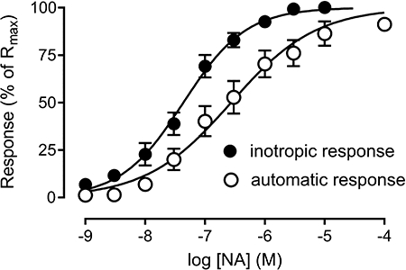
Experimental approach: The effects of adrenoceptor agonists on the rate of spontaneous contractions (automatic response) and the amplitude of electrically evoked contractions (inotropic response) were determined in left atria and ventricular myocytes isolated from Wistar rats.

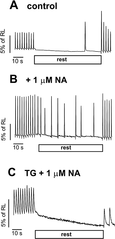
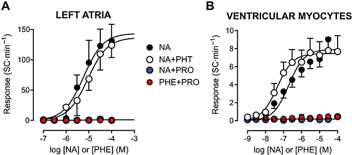
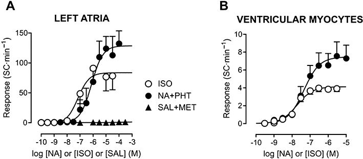
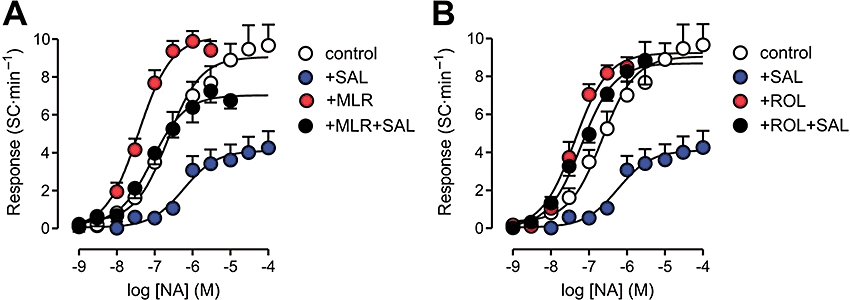
Key results: Catecholaminergic automatism was Ca(2+) -dependent, as it required a functional sarcoplasmic reticulum to be exhibited. Although both α- and β-adrenoceptor activation caused inotropic stimulation, only β(1) -adrenoceptors seemed to mediate the induction of spontaneous activity. Catecholaminergic automatism was enhanced and suppressed by β(2) -adrenoceptor blockade and stimulation respectively. Inhibition of either PDE3 or PDE4 (by milrinone and rolipram, respectively) potentiated the automatic response of myocytes to catecholamines. However, only rolipram abolished the attenuation of automatism produced by β(2) -adrenoceptor stimulation.

Conclusions and implications: α- and β(2) -adrenoceptors do not seem to be involved in the mediation of catecholaminergic stimulation of spontaneous activity in atrial and ventricular myocardium. However, a functional antagonism of β(1) - and β(2) -adrenoceptor activation was identified, the former mediating catecholaminergic myocardial automatism and the latter attenuating this effect. Results suggest that hydrolysis of cAMP by PDE4 is involved in the protective effect mediated by β(2) -adrenoceptor stimulation.


Figures

Figure 1
Figure 1
Records of ventricular myocyte shortening showing development of spontaneous contractions during interruption of electric stimulation (rest), in the absence (A) and presence (B and C) of noradrenaline (NA). Disabling sarcoplasmic reticulum function by thapsigargin (TG) treatment abolished spontaneous activity, even in the presence of noradrenaline (C). RL, resting cell length.
Figure 2
Figure 2
Inotropic (increase in systolic cell shortening) and automatic (rate of spontaneous contractions in the absence of electric stimulation) responses to noradrenaline (NA) determined in the same set of ventricular myocytes (n= 18). The responses were normalized to the respective maximum responses (Rmax). The mean pD2 value was significantly greater for the inotropic (7.36 ± 0.14) than for the automatic response (6.76 ± 0.16; P < 0.05).
Figure 3
Figure 3
Concentration–effect curves for adrenoceptor agonists in isolated rat left atria (A) and ventricular myocytes (B). The automatic response is expressed as the rate of spontaneous contractions (SC) during rest. Curves to noradrenaline (NA) were obtained in the absence of antagonists, as well as in the presence of the α- and β-adrenoceptor antagonists phentolamine (PHT, 1 µM) and propranolol (PRO, 1 µM) respectively. The response to the α-adrenoceptor agonist phenylephrine (PHE) was determined in the presence of PRO. Atropine (0.1 µM) was present throughout in all experiments with left atria. Values shown are means and SE. Curve parameters are shown in Table 1.
Figure 4
Figure 4
Concentration–effect curves for β-adrenoceptor agonists in isolated rat left atria (A) and ventricular myocytes (B). Curves are shown as in Figure 2. In both preparations, the maximum automatic response was greater for preferential activation of β1-adrenoceptors by noradrenaline (NA) under α-adrenoceptor blockade by phentolamine (PHT, 1 µM) than by non-selective β-adrenoceptor stimulation by isoprenaline (ISO). β2-adrenoceptor stimulation by salbutamol (SAL) in the presence of the β1-adrenoceptor antagonist metoprolol (MET, 0.5 µM) did not evoke automatism. All experiments with left atria were performed in the presence of 0.1 µM atropine. Values shown are means and SE. Curve parameters are shown in Table 1.
Figure 5
Figure 5
Concentration–effect curves for isoprenaline (ISO) and noradrenaline (NA) in isolated rat left atria (A) and ventricular myocytes (B–D). Curves were also determined during β2-adrenoceptor blockade with butoxamine (BUT, 0.3 µM) or ICI118551 (ICI, 0.1 µM), and in the presence of the β2-adrenoceptor agonist salbutamol (SAL, 10 µM). Experiments with left atria were performed in the presence of 0.1 µM atropine. Panels A and C show results obtained at 36°C, whereas the temperature was 23°C for experiments in panels B and D. Curve parameters are shown in Tables 1–3.
Figure 6
Figure 6
Concentration–effect curves for noradrenaline (NA) in isolated rat ventricular myocytes, determined in the presence and absence of the phosphodiesterase inhibitors milrinone (MLR, 3 µM, panel A) or rolipram (ROL, 3 µM, panel B). Some curves were determined during perfusion with the β2-adrenoceptor agonist salbutamol (SAL, 10 µM). Curve parameters are shown in Table 2.

Similar articles

Cited by

References

    1. Abi-Gerges A, Richter W, Lefebvre F, Mateo P, Varin A, Heymes C, et al. Decreased expression and activity of cAMP phosphodiesterases in cardiac hypertrophy and its impact on β-adrenergic cAMP signals. Circ Res. 2009;105:784–792. - PMC - PubMed
    1. Altschuld RA, Billman GE. β2-adrenoceptors and ventricular fibrillation. Pharmacol Ther. 2000;88:1–14. - PubMed
    1. Bassani RA, De Moraes S. Effects of repeated footshock stress on the chronotropic responsiveness of the isolated pacemaker of the rat: role of beta-2 adrenoceptors. J Pharmacol Exp Ther. 1988;246:316–321. - PubMed
    1. Bassani JWM, Yuan WL, Bers DM. Fractional SR Ca release is regulated by trigger Ca and SR Ca content. Am J Physiol. 1995;268:C1313–C1319. - PubMed
    1. Bassani RA, Bassani JWM, Lipsius SL, Bers DM. Diastolic Ca efflux in atrial pacemaker cells and Ca-overloaded myocytes. Am J Physiol. 1997;273:H886–H892. - PubMed

Publication types

MeSH terms

Substances