. 2011 Apr;162(7):1498-508.
doi: 10.1111/j.1476-5381.2010.01126.x.

Metformin inhibits HMGB1 release in LPS-treated RAW 264.7 cells and increases survival rate of endotoxaemic mice

Affiliations

Metformin inhibits HMGB1 release in LPS-treated RAW 264.7 cells and increases survival rate of endotoxaemic mice

Konstantin Tsoyi et al. Br J Pharmacol. 2011 Apr.

Abstract

Background and purpose: Recently, metformin, a well-known anti-diabetic drug, has been shown to possess anti-inflammatory activities. This study investigated the effect of metformin on the expression of pro-inflammatory cytokines including high mobility group box 1 (HMGB1) in lipopolysaccharide (LPS)-treated animals and cells.

Experimental approach: We investigated whether metformin inhibits the release of HMGB1 in LPS-treated RAW 264.7 cells and increases survival rate in endotoxaemic mice (lethal endotoxaemia was induced by an i.p. injection of LPS). This was achieved by a range of techniques including Western blotting, enzyme-linked immunosorbent assay, specific pharmacological inhibitors, knock out of α(1) -subunit of AMP-activated protein kinase (AMPK) and recombinant HMGB1.

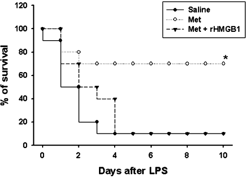
Key results: Both pre- and post-treatment with metformin significantly improved survival of animals during lethal endotoxaemia (survival rate was monitored up to 2 weeks), decreased serum levels of tumour necrosis factor-alpha (TNF-α), interleukin-1β, HMGB1 expression and myeloperoxidase activity in lungs. However, metformin failed to improve survival in endotoxaemic animals that had additionally been treated with recombinant HMGB1. In an in vitro study, metformin dose-dependently inhibited production of pro-inflammatory cytokines and HMGB1 release. Metformin activated AMPK by its phosphorylation. Compound C (pharmacological inhibitor of AMPK) and siAMPKα1 reversed the anti-inflammatory effect of metformin in LPS-treated cells.

Conclusions and implications: Our data indicate that metformin significantly attenuates the pro-inflammatory response induced by LPS both in vivo and in vitro. Metformin improved survival in a mouse model of lethal endotoxaemia by inhibiting HMGB1 release. AMPK activation was implicated as one of the mechanisms contributing to this inhibition of HMGB1 secretion.


Figures

Figure 1
Figure 1
Effect of metformin on the release of various cytokines, iNOS (NO) and COX-2 (PGE2) expression in LPS-activated macrophages. (A to E) Cells were pretreated with metformin (Met) 1, 5 and 10 mM for 1 h, then stimulated with LPS (1 µg·mL−1) for other 16 h. After incubation, culture medium samples were collected and subjected to elisas for IL-1β (A), TNF-α (B), IL-6 (C), PGE2 (E) and nitric oxide production by NO assay as described in Methods. Cells were harvested and iNOS and COX-2 protein levels were determined by Western blot. Data are presented as mean ± SD of three independent experiments. One-way analysis of variance was used to compare multiple group means followed by Newman–Keuls test (significance compared with control, **P < 0.01; significance compared with LPS, †P < 0.05 or ††P < 0.01). COX-2, cyclooxygenase-2; ELISA, enzyme-linked immunosorbent assay; IL-1β, interleukin-1β; IL-6, interleukin-6; iNOS, inducible nitric oxide synthase; PGE2, prostaglandin E2; TNF-α, tumour necrosis factor-alpha.
Figure 2
Figure 2
Effect of metformin on HMGB1 release in LPS-activated macrophages. (A and B) Cells were treated with metformin (1, 5 and 10 mM) for 1 h, then stimulated with LPS (1 µg·mL−1) for another 24 h. After incubation, culture medium was collected and subjected to HMGB1 anylasis (A). Cells were subjected to nuclear/cytosol fractionation and immunoblotted against HMGB1 as described in Methods. Blot bands are representative of three independent experiments. Data are presented as mean ± SD of three independent experiments. One-way analysis of variance was used to compare multiple group means followed by Newman–Keuls test (significance compared with control, **P < 0.01; significance compared with LPS, †P < 0.05 or ††P < 0.01). HMGB1, high mobility group box 1; LPS, lipopolysaccharides.
Figure 3
Figure 3
Metformin activates AMPK in macrophages. (A) Cells were treated with metformin (1, 2.5, 5, 10 mM) for 8 h. After incubation, phosphor- and total AMPK were assessed by Western blot. (B) Cells were incubated with metformin (5 mM) for 1, 2, 4, 8, and 12 h. After incubation, cells were lysed and subjected to Western blot for phosphor-AMPK and AMPK detection. Data are presented as mean ± SD of three independent experiments. One-way analysis of variance was used to compare multiple group means followed by Newman–Keuls test (significance compared with control, *P < 0.05; **P < 0.01). AMPK, AMP-activated protein kinase.
Figure 4
Figure 4
Pharmacological inhibition of AMPK reversed metformin-mediated anti-inflammatory effect in LPS-activated macrophages. Cells were treated with metformin (10 mM) in the presence or absence of compound C (comp C; 12 µM) for 1 h. Then cells were stimulated with LPS (1 µg·mL−1) for the next 24 h. After incubation, cells were lysed and subjected to Western blot (A); culture medium samples were extracted for HMGB1 detection by HMGB1 analysis (B) as described in Methods. Data are presented as mean ± SD of three independent experiments. One-way analysis of variance was used to compare multiple group means followed by Newman–Keuls test. Significance compared with control, **P < 0.01 and *P < 0.05; significance compared with LPS, ††P < 0.01; significance compare to Met (10 mM), §§P < 0.01. AMPK, AMP-activated protein kinase; HMGB1, high mobility group box 1; iNOS, inducible nitric oxide synthase; LPS, lipopolysaccharides.
Figure 5
Figure 5
Knock down of AMPK-α1 attenuates anti-inflammatory effect of metformin in LPS-activated macrophages. (A) Cells were transfected with scramble siRNA (ssiRNA, 100 nM) or with siAMPK-α1 (100 nM) and incubated for 16 h. After incubation, cells were harvested and AMPK or β-actin was detected by Western blot. (B and C) Cells were transfected with ssiRNA or siAMPK-α1. After transfection, cells were stimulated with LPS (1 µg·mL−1) in the presence or absence of metformin (10 mM). Cells were lysed, 24 h later, for iNOS detection. Culture medium was subjected to HMGB1 analysis as described in Methods. Data are presented as ±SD of three independent experiments. One-way analysis of variance was used to compare multiple group means followed by Newman–Keuls test. Significance compared with control, **P < 0.01 and *P < 0.05; significance compared with LPS, ††P < 0.01; significance compare to Met (10 mM), §§P < 0.01. AMPK, AMP-activated protein kinase; HMGB1, high mobility group box 1; iNOS, inducible nitric oxide synthase; LPS, lipopolysaccharides.
Figure 6
Figure 6
Metformin improves survival in lipopolysaccharides (LPS)-induced mouse model of sepsis. (A) BALB/c mice were pretreated with either saline (i.p., n= 20), metformin (50 mg·kg−1, i.p., n= 20) or metformin (100 mg·kg−1, i.p., n= 20) 2 h prior to LPS challenge (15 mg·kg−1, i.p.). At 12, 24, 48, 72 and 96 h after the onset of endotoxaemia, animals were administered (i.p.) either saline or metformin 50 mg·kg−1 or 100 mg·kg−1. (B) BALB/c mice were subjected to lethal endotoxaemia (LPS, 15 mg·kg−1, i.p.) and 12 h later treated with either saline (i.p., n= 20), metformin (50 mg·kg−1, i.p., n= 20) or metformin (100 mg·kg−1, i.p., n= 20). Treatments were repeated at 24 h, 48 h, 72 h, and 96 h after induction of endotoxaemia. Survival was monitored daily, for up to two weeks. The Kaplan–Meier program was utilized to compare the differences in mortality rates between groups. Significance compared with saline, *P < 0.05.
Figure 7
Figure 7
Metformin attenuates inflammation and neutrophil infiltration into lungs induced by LPS. BALB/c mice were treated with either saline (i.p., n= 5); saline (i.p.) +LPS (15 mg·kg−1, i.p.) (n= 5) or metformin (100 mg·kg−1) +LPS (15 mg·kg−1, i.p.) (n= 5); 24 h after LPS treatment mice were anaesthetized, blood and lungs were extracted. Serum levels of IL-1β and TNF-α were detected by elisa, HMGB1 by HMGB1 analysis. Lungs were homogenized and MPO activity was measured by elisa. Data are presented as mean ± SD of three independent experiments. One-way analysis of variance was used to compare multiple group means followed by Newman–Keuls test (significance compared with control, *P < 0.05 or **P < 0.01; significance compared with LPS, †P < 0.05 or ††P < 0.01). IL-1β, interleukin-1 β; LPS, lipopolysaccharide; MPO, myeloperoxidase; TNF-α, tumour necrosis factor α.
Figure 8
Figure 8
rHMGB1 reversed the therapeutic effect of metformin on lethal endotoxaemia. BALB/c mice were pretreated with saline (i.p., n= 10), metformin (100 mg·kg−1, i.p., n= 10) 2 h prior to LPS injection (15 mg·kg−1, i.p.). At 12, 24, 48, 72 and 96 h after the onset of endotoxaemia, animals were administered (i.p.) saline or metformin (100 mg·kg−1) + rHMGB1 (100 µg). Survival was monitored daily, for up to 2 weeks. The Kaplan–Meier program was utilized to compare the differences in mortality rates between groups. Significance compared with saline, *P < 0.05. HMGB1, high mobility group box 1; rHMGB1, recombinant human high mobility group box 1.

Similar articles

Cited by

References

    1. Abdulahad DA, Westra J, Limburg PC, Kallenberg CG, Bijl M. HMGB1 in Systemic Lupus Erythematosus: its role in cutaneous lesions development. Autoimmun Rev. 2010;9:661–665. [Epub ahead of print] - PubMed
    1. Andersson U, Harris HE. The role of HMGB1 in the pathogenesis of rheumatic disease. Biochim Biophys Acta. 2010;1799:141–148. - PubMed
    1. Andersson U, Wang H, Palmblad K, Aveberger AC, Bloom O, Erlandsson-Harris H, et al. High mobility group 1 protein (HMG-1) stimulates proinflammatory cytokine synthesis in human monocytes. J Exp Med. 2000;192:565–570. - PMC - PubMed
    1. Bailey CJ, Turner RC. Metformin. N Engl J Med. 1996;334:574–579. - PubMed
    1. Bergheim I, Luyendyk JP, Steele C, Russell GK, Guo L, Roth RA, et al. Metformin prevents endotoxin-induced liver injury after partial hepatectomy. J Pharmacol Exp Ther. 2006;316:1053–1061. - PubMed

Publication types

MeSH terms