. 2010 Nov 19:11:87.
doi: 10.1186/1471-2199-11-87.

Translational independence between overlapping genes for a restriction endonuclease and its transcriptional regulator

Affiliations

Translational independence between overlapping genes for a restriction endonuclease and its transcriptional regulator

Meenakshi K Kaw et al. BMC Mol Biol. .

Abstract

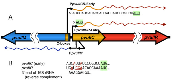
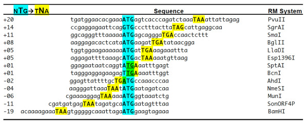
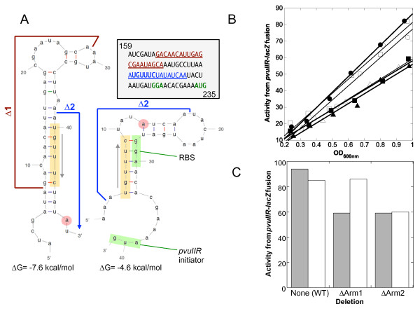
Background: Most type II restriction-modification (RM) systems have two independent enzymes that act on the same DNA sequence: a modification methyltransferase that protects target sites, and a restriction endonuclease that cleaves unmethylated target sites. When RM genes enter a new cell, methylation must occur before restriction activity appears, or the host's chromosome is digested. Transcriptional mechanisms that delay endonuclease expression have been identified in some RM systems. A substantial subset of those systems is controlled by a family of small transcription activators called C proteins. In the PvuII system, C.PvuII activates transcription of its own gene, along with that of the downstream endonuclease gene. This regulation results in very low R.PvuII mRNA levels early after gene entry, followed by rapid increase due to positive feedback. However, given the lethal consequences of premature REase accumulation, transcriptional control alone might be insufficient. In C-controlled RM systems, there is a ± 20 nt overlap between the C termination codon and the R (endonuclease) initiation codon, suggesting possible translational coupling, and in many cases predicted RNA hairpins could occlude the ribosome binding site for the endonuclease gene.

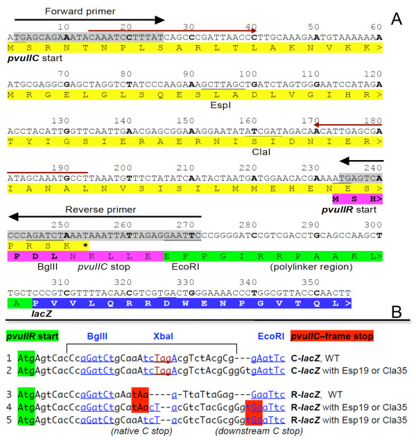
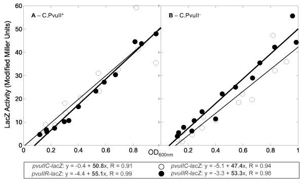
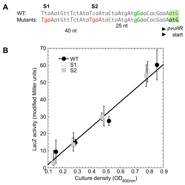
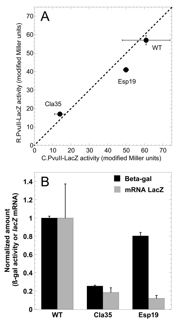
Results: Expression levels of lacZ translational fusions to pvuIIR or pvuIIC were determined, with the native pvuII promoter having been replaced by one not controlled by C.PvuII. In-frame pvuIIC insertions did not substantially decrease either pvuIIC-lacZ or pvuIIR-lacZ expression (with or without C.PvuII provided in trans). In contrast, a frameshift mutation in pvuIIC decreased expression markedly in both fusions, but mRNA measurements indicated that this decrease could be explained by transcriptional polarity. Expression of pvuIIR-lacZ was unaffected when the pvuIIC stop codon was moved 21 nt downstream from its WT location, or 25 or 40 bp upstream of the pvuIIR initiation codon. Disrupting the putative hairpins had no significant effects.

Conclusions: The initiation of translation of pvuIIR appears to be independent of that for pvuIIC. Direct tests failed to detect regulatory rules for either gene overlap or the putative hairpins. Thus, at least during balanced growth, transcriptional control appears to be sufficiently robust for proper regulation of this RM system.


Figures

Figure 1
Figure 1
PvuII transcripts. A. The diagram indicates the expected transcripts early or later after the PvuII genes enter a new host cell. The timing is inferred from in vivo transcript mapping in the presence or absence of active C.PvuII [7], and following synchronous infection of cells with bacteriophage carrying the PvuII genes [28]. "C boxes" are the binding sites for the autogenously-activating C protein. The two pvuIIM (methyltransferase) promoters appear to be constitutive. B. The ribosome-binding (Shine-Dalgarno) sequences are predicted based on location relative to the pvuIIC start codon (shaded green), and comparison to the Logo for ribosome binding sites adapted from [32].
Figure 2
Figure 2
Relative Locations of C Terminators and R Initiators of Translation. Selected C.PvuII orthologs that are upstream of known or candidate restriction endonuclease genes were aligned via the endonuclease initiation codons to illustrate the range of relative positions. Names and gene boundaries are available at REBase [1]. Numbers at the left are center-to-center distances between the C gene termination codon and the R gene initiation codon.
Figure 3
Figure 3
pvuII-lacZ transcriptional fusions. A. Sequence from the pvuIIC initiator codon to the pvuIIR-fused lacZ gene. The vector, and source of the polylinker (green) and lacZ gene (blue), is pLex3B (ATCC #87200) [58]. The primers indicated by black arrows and gray shading were used to PCR-amplify pvuIIC and part of pvuIIR; pvuIIR retains its native RBS. The pvuIIC gene includes two unique sites, ClaI and EspI (equivalent to BlpI), at which different null mutations in pvuIIC were generated as previously described [17]. The WT and three different mutants were cloned between the vector XmnI and EcoRI sites such that 'A' of the XmnI site blunt ligated to the 'TG' of the insert on the 5' end to regenerate the pvuIIC initiation codon (under the control of the vector's promoter and RBS); the 3' end ligation used the EcoRI site. In derivatives, synthetic oligonucleotides were inserted between the BglII and EcoRI sites (underlined) to fuse pvuIIC to lacZ or to introduce other changes. The pair of red arrows indicates primers used for mRNA quantitation. B. Oligonucleotides used to alter the pvuIIC-pvuIIR overlap region. A 30 bp oligonucleotide was cloned between the BglII and EcoRI sites (in the sequence shown in Figure 3) to put pvuIIC in-frame with the lacZ gene. In each case, the pvuIIR initiation codon is highlighted in green, and the pvuIIC-frame terminator in red. A unique XbaI site was included to help identify the desired clones and to facilitate shifting the fusion reading frame. A pvuIIC stop codon was introduced in some cases (bottom sequences), to restore it to its native location relative to the pvuIIR initiator. In lines 1 and 2, the pvuIIC-frame terminator is off to the right (not shown).
Figure 4
Figure 4
Expression of pvuIIC-lacZ and pvuIIR-lacZ translational fusions in the presence and absence of C.PvuII. Cultures of E. coli TOP10 were grown in exponential phase in defined rich medium containing IPTG, and samples were taken at several times for β-galactosidase assays. If the cells are in balanced growth, the plot of activity vs. culture density should be linear. The translational fusions were to pvuIIC (open circles) or pvuIIR (closed circles), and in both cases are transcribed from a C-independent vector promoter. The equations resulting from linear regression are shown. A. A compatible plasmid is providing active C.PvuII in trans. B. As in (A), except the plasmid is providing an inactive version of C.PvuII.
Figure 5
Figure 5
Stop codons created in pvuIIC upstream of pvuIIR-lacZ fusion initiation site. The native pvuIIC translation terminator is downstream of the pvuIIR initiation codon (see top line of Figure 2). A. To test for translational coupling, site-directed mutation was used to introduce pvuIIC terminators farther upstream. The pvuIIR RBS and intiation codon are indicated in green. The two introduced stop codons were in two independent clones. B. lacZ activity of the WT (filled circles) and the mutants (open squares have terminator indicated "S1" in part A; filled squares represent S2). The linear fit is to the WT data.
Figure 6
Figure 6
Effects of pvuIIC mutation. A. Comparison of pvuIIC and pvuIIR translation with WT and mutant variants of pvuIIC. The slopes from triplicate experiments such as that shown in Figure 4 are plotted, showing standard errors (where bars are not visible, errors were smaller than the symbol). In this correlogram, activity from the pvuIIC-lacZ translational fusion is shown on the x-axis, and that from the pvuIIR-lacZ fusion of the same mutant is on the y-axis; if the two fusions for a given variant have equal translation, the point would fall on the dotted line. B. Effects of mutation on mRNA levels and translation activity of pvuIIR-lacZ fusions. Reporter pvuIIR fusions with WT pvuIIC upstream, or with the Cla35 (frameshift) or Esp19 (in-frame) pvuIIC mutations, were grown in triplicate. Real-time RT-PCR was carried out as described in Methods, using the SYBR green method [60] and primers specific to lacZ. Quantitation was based on a standard curve with normalization to recA mRNA. Amounts of mRNA (gray bars) are normalized to the level in the strain carrying WT pvuIIC. The black bars indicate lacZ activity measurements in the same cultures, measured as shown in Figure 4 and normalized to the WT value. Standard errors are shown.
Figure 7
Figure 7
RNA secondary structure upstream of pvuIIR. A. Putative alternative hairpins in pvuIICR mRNA. The sequence from the pvuIIC gene just upstream of pvuIIR is shown, with numbering corresponding to that in Figure 3A. As previously described [9], the program MFOLD [61] predicts alternative hairpin structures, the downstream one of which would occlude the RBS (structure on right, green highlight). The orange highlighting shows a sequence shared between the two structures, making them mutually exclusive. The red circle indicates the position of a termination product in previous primer extension reactions [7]. The boundaries of two in-frame deletions are indicated. B. Effect of in-frame hairpin arm deletions on translation of pvuIIR. The deletions shown in (A) were introduced into the pvuIIR-lacZ translational fusion, and β-galactosidase activity was measured as described for Figure 4. This was repeated in the presence of WT (closed symbols, gray bars) or null mutant forms of C.PvuII (open symbols, white bars) provided in trans from compatible plasmids. Plot of activity vs. culture density, for WT (circles), arm 1 deletion mutant (squares), or arm 2 deletion mutant (triangles). C. Slopes from (B) are shown to facilitate comparison.

Similar articles

Cited by

References

    1. Roberts RJ, Vincze T, Posfai J, Macelis D. REBASE--a database for DNA restriction and modification: enzymes, genes and genomes. Nucleic Acids Res. 2010. pp. D234–236. - DOI - PMC - PubMed
    1. Releigh EA, Brooks JE. Restriction Modification systems: where they are and what they do. Chapman & Hall, New York, N.Y; 1998.
    1. Kobayashi I. Behavior of restriction-modification systems as selfish mobile elements and their impact on genome evolution. Nucleic Acids Res. 2001;29(18):3742–3756. doi: 10.1093/nar/29.18.3742. - DOI - PMC - PubMed
    1. Ichige A, Kobayashi I. Stability of EcoRI restriction-modification enzymes in vivo differentiates the EcoRI restriction-modification system from other postsegregational cell killing systems. J Bacteriol. 2005;187(19):6612–6621. doi: 10.1128/JB.187.19.6612-6621.2005. - DOI - PMC - PubMed
    1. Nakayama Y, Kobayashi I. Restriction-modification gene complexes as selfish gene entities: roles of a regulatory system in their establishment, maintenance, and apoptotic mutual exclusion. Proc Natl Acad Sci USA. 1998;95(11):6442–6447. doi: 10.1073/pnas.95.11.6442. - DOI - PMC - PubMed

Publication types

Substances