Review
. 2010 Nov 22:5:30.
doi: 10.1186/1750-1172-5-30.

Fabry disease

Affiliations
Review

Fabry disease

Dominique P Germain. Orphanet J Rare Dis. .

Abstract

Fabry disease (FD) is a progressive, X-linked inherited disorder of glycosphingolipid metabolism due to deficient or absent lysosomal α-galactosidase A activity. FD is pan-ethnic and the reported annual incidence of 1 in 100,000 may underestimate the true prevalence of the disease. Classically affected hemizygous males, with no residual α-galactosidase A activity may display all the characteristic neurological (pain), cutaneous (angiokeratoma), renal (proteinuria, kidney failure), cardiovascular (cardiomyopathy, arrhythmia), cochleo-vestibular and cerebrovascular (transient ischemic attacks, strokes) signs of the disease while heterozygous females have symptoms ranging from very mild to severe. Deficient activity of lysosomal α-galactosidase A results in progressive accumulation of globotriaosylceramide within lysosomes, believed to trigger a cascade of cellular events. Demonstration of marked α-galactosidase A deficiency is the definitive method for the diagnosis of hemizygous males. Enzyme analysis may occasionnally help to detect heterozygotes but is often inconclusive due to random X-chromosomal inactivation so that molecular testing (genotyping) of females is mandatory. In childhood, other possible causes of pain such as rheumatoid arthritis and 'growing pains' must be ruled out. In adulthood, multiple sclerosis is sometimes considered. Prenatal diagnosis, available by determination of enzyme activity or DNA testing in chorionic villi or cultured amniotic cells is, for ethical reasons, only considered in male fetuses. Pre-implantation diagnosis is possible. The existence of atypical variants and the availability of a specific therapy singularly complicate genetic counseling. A disease-specific therapeutic option - enzyme replacement therapy using recombinant human α-galactosidase A - has been recently introduced and its long term outcome is currently still being investigated. Conventional management consists of pain relief with analgesic drugs, nephroprotection (angiotensin converting enzyme inhibitors and angiotensin receptors blockers) and antiarrhythmic agents, whereas dialysis or renal transplantation are available for patients experiencing end-stage renal failure. With age, progressive damage to vital organ systems develops and at some point, organs may start to fail in functioning. End-stage renal disease and life-threatening cardiovascular or cerebrovascular complications limit life-expectancy of untreated males and females with reductions of 20 and 10 years, respectively, as compared to the general population. While there is increasing evidence that long-term enzyme therapy can halt disease progression, the importance of adjunctive therapies should be emphasized and the possibility of developing an oral therapy drives research forward into active site specific chaperones.


Figures

Figure 1
Figure 1
Angiokeratoma: the angiokeratoma are small, raised, dark-red spots that increase in number and size with age and can occur singly or in clusters. They are typically found on the lower back (A), buttocks (C), groin, flanks (D) and upper thighs but their distribution may be restricted to a limited area, such as the umbilicus (B).
Figure 2
Figure 2
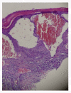
Skin biopsy (light microscopy): histologically, the typical skin lesion is a small superficial angioma caused by cumulative damage of the vascular cells of the dermis with vessel dilation. Courtesy: Dr Juan M. POLITEI, Buenos Aires, Argentina.
Figure 3
Figure 3
Kidney biopsy (light microscopy): low power view of a glomerulus on a core needle biopsy in Fabry disease, × 320. Courtesy Pr Marie-Claire GUBLER, Paris, France
Figure 4
Figure 4
Kidney biopsy (light microscopy): the purple stain is on the podocytes where there is the most prominent collection of Gb3 in the kidney. Courtesy Pr Laura BARISONI, New-York University, New York, USA.
Figure 5
Figure 5
Kidney biopsy: electron microscopy shows massive storage of glycosphingolipids in the lysosomes of podocytes. Courtesy: Pr Marie-Claire GUBLER, Paris, France.
Figure 6
Figure 6
Kidney biopsy (electron microscopy): glycosphingolipid inclusions of various size and shape are seen in the cells of distal tubules of the kidney in Fabry disease.
Figure 7
Figure 7
Kidney biopsy (electron microscopy): glycolipid inclusions in the endothelial and smooth muscle cells of a renal arteriole. No storage can be seen in the proximal tubule (TP), × 8200. Courtesy: Pr Marie-Claire GUBLER, Paris, France.
Figure 8
Figure 8
Echocardiography: parasternal long axis showing diffuse left ventricular hypertrophy with increased septal thickness. Courtesy: Pr Albert A. HAGEGE, University René Descartes, Paris, France.
Figure 9
Figure 9
Echocardiography: parasternal short axis showing left ventricular hypertrophy. Courtesy: Pr Albert A. HAGEGE, Université René Descartes, Paris, France.
Figure 10
Figure 10
Cardiac MRI for the assessment of left ventricular hypertrophy and fibrosis: A: Left ventricular hypertrophy in a 51-year-old male patient with cerebrovascular involvement and end stage renal disease (dialysis). B: Hypertrophic cardiomyopathy in a 56-year-old male patient with arrythmya, leukoareiosis and kidney transplant. C: Late enhancement after gadolinium in a 63-year-old female patient with end stage renal disease (dialysis).
Figure 11
Figure 11
Tissue Doppler of the mitral annulus: near normal systolic function. Courtesy: Pr Albert A. HAGEGE, University René Descartes, Paris, France.
Figure 12
Figure 12
Doppler: near normal systolic function (same patient as in figure 10). Courtesy: Pr Albert A. HAGEGE, University René Descartes, Paris, France.
Figure 13
Figure 13
ECG: showing electrical signs of left ventricular hypertrophy with increased Sokolow index, depressed ST segment and negative T waves in left derivations
Figure 14
Figure 14
24-hour-ECG holter: is recommended at baseline and during follow-up of enzyme replacement therapy if arrhythmia is suspected on ECG or palpitations are reported by the patient.
Figure 15
Figure 15
Aortic root dilatation: echocardiography shows aortic root diameter of 47 mm in a 51-year-old male patient with Fabry disease. Courtesy: Pr Olivier DUBOURG and Pr Dominique GERMAIN, University of Versailles - St Quentin en Yvelines (UVSQ), Versailles, France.
Figure 16
Figure 16
Aortic root dilatation in a patient suffering from Fabry disease: magnetic resonance imaging (MRI) showing aortic root dilatation in Fabry disease. Pr Dominique GERMAIN, University of Versailles - St Quentin en Yvelines (UVSQ), Versailles, France
Figure 17
Figure 17
Stroke in a patient affected with Fabry disease: axial brain MRI section showing stroke of the left cerebellar hemisphere that revealed Fabry disease in an otherwise asymptomatic 27-year-old male patient.
Figure 18
Figure 18
Dolichoectasia of the vertebro-basilar circulation: time of flight magnetic resonance angiographies showing ectatic vessels in four patients affected with Fabry disease.
Figure 19
Figure 19
Cerebral white matter hyperintensities, lacuna and microbleeds: A. Fluid-attenuated inversion recovery (FLAIR)-weighted axial MRI section showing multiple white matter lesions in the cerebral hemispheres in a 53-year-old male patient who had a Fazekas score of 9. B. Lacuna and microbleeds in the same patient. Courtesy: Dr Robert CARLIER and Dr Frédéric COLAS, CHU Raymond Poincaré, Garches, France.
Figure 20
Figure 20
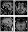
The pulvinar sign: T1-weighted sagital (A) and axial (B) MRI sections showing the pulvinar sign in a 66 year-old male patient. T1-weighted sagital (C) and axial (D) MRI section showing symmetrical high signals in the pulvinar region in a 42-year-old male patient. Courtesy: Dr Robert CARLIER and Dr Frédéric COLAS, CHU Raymond Poincaré, Garches, France.
Figure 21
Figure 21
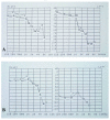
Hypoacousia in patients affected with Fabry disease: A. Hypoacousia in a 39-year-old male with hypertrophic cardiomyopathy, cerebral lacuna and kidney transplant. B. Sudden deafness of the left ear and bilateral hypoacousia in a 54-year-old male patient with tinnitus, vertigo, vertebro-basilar TIA, hypertrophic cardiomyopathy and kidney transplant. Courtesy: Dr Philippe AUBERT and Dr Karelle BENISTAN, CHU Raymond Poincaré, Garches, France.
Figure 22
Figure 22
Cornea of a female patient heterozygote for FD: sub-epithelial brown lines show the typical pattern of so-called "cornea verticillata". These opacities do not impair the visual acuity. Courtesy: Dr Juan-Manuel POLITEI, Buenos-Aires, Argentina.
Figure 23
Figure 23
Standard Goldman visual field of the left eye of a patient affected with Fabry disease: the blind spot is enlarged. Courtesy: Dr Christophe ORSSAUD, Paris, France.
Figure 24
Figure 24
Dual-energy X-ray absorptiometry (DEXA) assessment of bone mineral density of the femoral neck (A) and the lumbar spine (B): T scores of - 4.2 and - 4.3 were found at the hip (A) and lumbar spine (B), respectively in a 53 year-old male patient affected with Fabry disease. Courtesy: Dr Caroline LEBRETON, CHU Raymond Poincaré, Garches, France.
Figure 25
Figure 25
Bone magnetic resonance imaging in a Fabry patient with severe osteoporosis: A (STIR, sagittal view) and B (T1, sagital median): several vertebral body fractures are seen, without signal anomaly in T1 or T2 in favor of ancient fractures. A mild spondylolisthesis of L5 on S1 can be observed. C (T2, axial view): fracture of the right pedicula of L5 (arrow) in a 72-year-old patient with severe osteoporosis. Courtesy: Dr Robert CARLIER, CHU Raymond Poincaré, Garches, France.
Figure 26
Figure 26
Genotyping of the GLA gene in heterozygous females: A. Patient CB, a 17-year-old girl, was shown to carry a T to G transversion in exon 6 at position 884 in the cDNA sequence. This nucleotide substitution alters the codon (TTC) for phenylalanine to the codon (TGC) for cysteine at position 295 of the α-galactosidase A protein (p.Phe295Cys). B. Patient ZB, a 46-year-old woman, was shown to carry a T to G transversion in exon 1 at position 125 in the cDNA sequence. This nucleotide substitution alters the codon (ATG) for methionine to the codon (AGG) for arginine at position 42 of the α-galactosidase A protein (p.Met42Arg). C. Patient NL, a 63-year-old woman was shown to carry a G to T transversion in exon 6 at position 982 in the cDNA sequence. This nucleotide substitution alters the codon (GGG) for glycine to the codon (TGG) for tryptophan at position 328 of the α-galactosidase A protein (p.Gly328Trp). Despite scanning of the rest of the gene, no other sequence abnormality was found. Courtesy: Pr Xavier JEUNEMAITRE and Dr Anne-Laure FAURET, HEGP, Paris, France.
Figure 27
Figure 27
Sequencing of PCR products obtained from amplification of DNA directly eluted from a 3-mm punch of dried blood spot (DBS) on filter paper: a 60-year-old man with left ventricular hypertrophy of unknown origin was enrolled in a screening protocol for FD. Markedly decreased α-galactosidase activity was found on DBS. Using a second DBS, the patient was subsequently shown to carry a T to C transition in exon 2 at position 337 in the cDNA sequence of the GLA gene (c.337T > C). This nucleotide substitution alters the codon (TTT) for phenylalanine to the codon (CTT) for leucine at position 113 of the α-galactosidase A protein (p.Phe113Leu). Pr Dominique GERMAIN, University of Versailles - St Quentin en Yvelines (UVSQ), Versailles, France
Figure 28
Figure 28
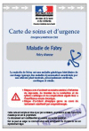
Emergency healthcare card from the French Ministry of Health: an emergency healthcare card was created by the Ministry of Health, the center of excellence for Fabry disease and patients' associations for Fabry disease or lysosomal storage diseases. The card is made of two parts: one of which contains general data on FD while the second one includes the personal medical history and medications of the patient in order to provide useful information for emergency care situations.
Figure 29
Figure 29
Median estimated glomerular filtration rate (eGFR; ml/min per 1.73 m2) over time in 44 patients treated with agalsidase beta for 54 months: Patients in the “as treated” population maintained a stable median eGFR during the 54-month treatment. Subgroup analyses of patients who were stratified by baseline proteinuria (>1 g/24 h versus <1 g/24 h) showed differences in the rate of eGFR decline during the 54-mo treatment period [309]. High (>1 g/24 h) baseline proteinuria was associated with higher rate of eGFR decline and increased probability of renal events.
Figure 30
Figure 30
Long-term agalsidase beta therapy decreases Gb3 accumulation in podocytes: A) Kidney biopsy which was obtained prior to agalsidase beta therapy shows dark-staining granules in podocytes. B) By month 54, fewer Gb3 inclusions are evident from a specimen which was obtained from the same patient. Methylene blue/azure II stain, magnification × 400 [309].
Figure 31
Figure 31
Skin rash during infusion of recombinant α-galactosidase A in a patient with positive IgE antibodies to agalsidase beta: In year 2002, a 39-year-old male Fabry patient (GLA mutation p.Ala121Pro) was initially treated with agalsidase beta (1 mg/kg EOW). ERT was changed to agalsidase alfa (0.2 mg/kg EOW) after 18 months due to poor tolerance (mild laryngeal edema, urticaria and chills during infusions). Two years later, a rash appeared on both arms during agalsidase alfa infusions. In 2007, concomittant deterioration of kidney function on agalsidase alfa (mGFR decreased from 85 to 70 mL/min/1.73 m2) led to switch ERT back to agalsidase beta. No data was obtained with respect to antibodies (IgG or IgE) to agalsidase alfa. After 1 year of agalsidase beta therapy, extensive skin rash and bronchospasm appeared during the infusions despite premedication (hydroxyzine, paracetamol and oral steroids) and minimal infusion rates (0.05 - 0.2 mg/min) and kidney function kept on deteriorating (mGFR = 54 mL/min/1.73 m2). The patient tested positive for IgE to agalsidase beta and ERT was discontinued. Mutation p.Ala121Pro is not responsive to the ASSC deoxygalactonojirymicin [424]. Both rechallenge protocol and concomitant use of immunosuppressive therapy and ERT are currently being considered.
Figure 32
Figure 32
Proposed mechanism of action of active site-specific chaperones (ASSCs): A: During synthesis of a wild-type lysosomal enzyme, cells assemble amino acids in a correctly folded tertiary structure. Molecular chaperones are naturally occurring molecules that assist in protein folding. B: In contrast, mutant misfolded lysosomal enzymes are unstable and retained in the endoplasmic reticulum (ER) where they may not meet quality control and are prone to endoplasmic reticulum-associated degradation (ERAD). C: ASSCs are designed to stabilize and rescue misfolded lysosomal enzymes, leading to reduced ER retention or accumulation, and enhanced trafficking to the Golgi apparatus and the lysosome where they dissociate from the enzyme.

References

    1. Anderson W. A case of "Angeio-keratoma". Br J Dermatol. 1898;10:113–117. doi: 10.1111/j.1365-2133.1898.tb16317.x. - DOI
    1. Fabry J. Ein Beitrag zur Kenntnis der Purpura haemorragica nodularis (Purpura papulosa hemorrhagica Hebrae) Arch Dermatol Syphilol. 1898;43:187–200. doi: 10.1007/BF01986897. - DOI
    1. Sweeley CC, Klionsky B. Fabry's disease: classification as a sphingolipidosis and partial characterization of a novel glycolipid. J Biol Chem. 1963;238:3148–3150. - PubMed
    1. Brady RO, Gal AE, Bradley RM, Martensson E, Warshaw AL, Laster L. Enzymatic defect in Fabry's disease: ceramide-trihexosidase deficiency. N Engl J Med. 1967;276:1163–1167. doi: 10.1056/NEJM196705252762101. - DOI - PubMed
    1. Kint JA. The enzyme defect in Fabry's disease. Nature. 1970;227:1173. doi: 10.1038/2271173b0. - DOI - PubMed

Publication types

MeSH terms

Substances