

Leiomyosarcoma of intravascular origin--a rare tumor entity: clinical pathological study of twelve cases
- PMID: 21092216
- PMCID: PMC3012034
- DOI: 10.1186/1477-7819-8-103
Leiomyosarcoma of intravascular origin--a rare tumor entity: clinical pathological study of twelve cases
Abstract
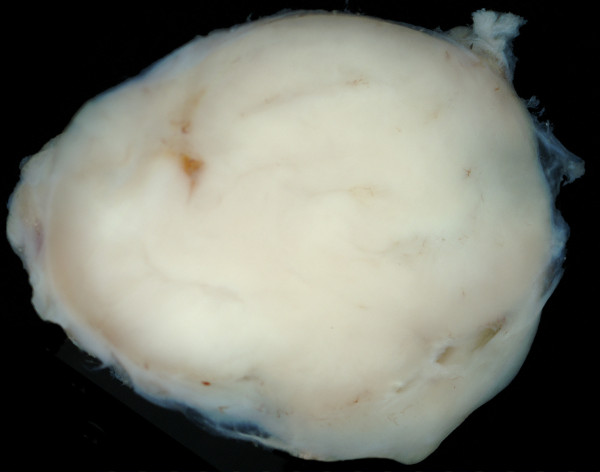
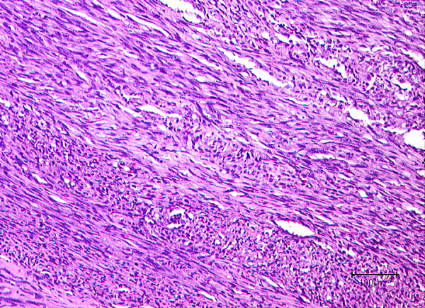
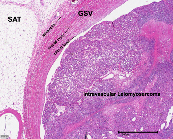
Background: Leiomysarcoma of intravascular origin is an exceedingly rare entity of malignant soft tissue tumors. They are most frequently encountered in the retroperitoneum arising from the inferior vena cava and are scarcely found to arise from vessels of the extremities. These tumors were analysed with particular reference to treatment outcome and prognosis. The aim of this article is to broaden the knowledge of the clinical course of this rare malignancy.
Method: During 2000 and 2009 twelve patients were identified with an intravascular origin of a leiomyosarcoma. Details regarding the clinical course, follow-up and outcome were assessed with focus on patient survival, tumor relapse and metastases and treatment outcome. 3 year survival probability was calculated using Kaplan-Meier method.
Results: Vascular leiomyosarcomas accounted for 0.7% of all malignant soft tissue tumors treated at our soft tissue sarcoma reference center. The mean follow up period was 38 months. Tumor relapse was encountered in six patients. 6 patients developed metastatic disease. The three year survival was 57%.
Conclusion: Vascular leiomysarcoma is a rare but aggressive tumor entity with a high rate of local recurrence and metastasis.
Figures
References
-
- Issels R. Manual Knochentumoren und Weichteilsarkome. 4. München: W Zuckerschwerdt Verlag; 2004.
-
- Weiss SWGJ. In: (Hrsg) Enzinger and Weiss's soft tissue tumors. 4. Weiss SW, Goldblum JR, editor. Mosby, StLouis Baltimore Berlin; 2001. Leiomysarcoma; pp. 727–748.
MeSH terms
LinkOut - more resources
Full Text Sources
