Comparative Study
. 2010;14(6):R209.
doi: 10.1186/cc9332. Epub 2010 Nov 23.

Validation of a new transpulmonary thermodilution system to assess global end-diastolic volume and extravascular lung water

Affiliations
Comparative Study

Validation of a new transpulmonary thermodilution system to assess global end-diastolic volume and extravascular lung water

Karim Bendjelid et al. Crit Care. 2010.

Abstract

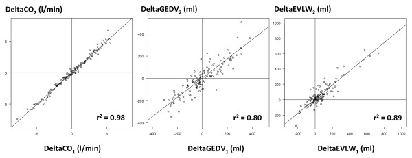
Introduction: A new system has been developed to assess global end-diastolic volume (GEDV), a volumetric marker of cardiac preload, and extravascular lung water (EVLW) from a transpulmonary thermodilution curve. Our goal was to compare this new system with the system currently in clinical use.

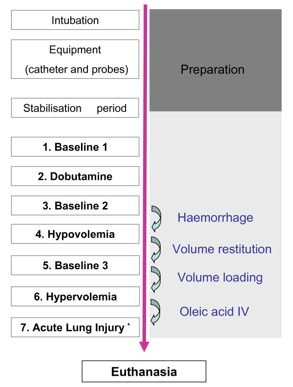
Methods: Eleven anesthetized and mechanically ventilated pigs were instrumented with a central venous catheter and a right (PulsioCath; Pulsion, Munich, Germany) and a left (VolumeView™; Edwards Lifesciences, Irvine, CA, USA) thermistor-tipped femoral arterial catheter. The right femoral catheter was used to measure GEDV and EVLW using the PiCCO(2)™ (Pulsion) method (GEDV(1) and EVLW(1), respectively). The left femoral catheter was used to measure the same parameters using the new VolumeView™ (Edwards Lifesciences) method (GEDV(2) and EVLW(2), respectively). Measurements were made during inotropic stimulation (dobutamine), during hypovolemia (bleeding), during hypervolemia (fluid overload), and after inducing acute lung injury (intravenous oleic acid).

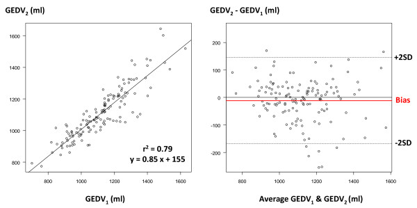
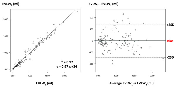
Results: One hundred and thirty-seven paired measurements were analyzed. GEDV(1) and GEDV(2) ranged from 701 to 1,629 ml and from 774 to 1,645 ml, respectively. GEDV(1) and GEDV(2) were closely correlated (r(2) = 0.79), with mean bias of -11 ± 80 ml and percentage error of 14%. EVLW(1) and EVLW(2) ranged from 507 to 2,379 ml and from 495 to 2,222 ml, respectively. EVLW(1) and EVLW(2) were closely correlated (r(2) = 0.97), with mean bias of -5 ± 72 ml and percentage error of 15%.

Conclusions: In animals, and over a very wide range of values, a good agreement was found between the new VolumeView™ system and the PiCCO™ system to assess GEDV and EVLW.


Figures

Figure 1
Figure 1
Transpulmonary thermodilution curve. The assessment of global end-diastolic volume (GEDV) by the PiCCO™ system is based on the mean transit time (MTt) and exponential downslope time (DSt), while the assessment of GEDV by the new VolumeView™ method is based on MTt, maximum ascending slope (S1) and maximum descending slope (S2).
Figure 2
Figure 2
Flow chart of the experimental protocol. *Multiple measurements. IV, intravenous.
Figure 3
Figure 3
Cardiac output comparison. Left: correlation between cardiac output (CO) measured by the PiCCO™ system (CO1) and the VolumeView™ system (CO2). Right: Bland-Altman representation depicting the agreement between both methods. SD, standard deviation.
Figure 4
Figure 4
Global end-diastolic volume comparison. Left: correlation between global end-diastolic volume (GEDV) measured by the PiCCO™ system (GEDV1) and the VolumeView™ system (GEDV2). Right: Bland-Altman representation depicting the agreement between both methods. SD, standard deviation.
Figure 5
Figure 5
Extravascular lung water comparison. Left: correlation between extravascular lung water (EVLW) measured by the PiCCO™ system (EVLW1) and the VolumeView™ system (EVLW2). Right: Bland-Altman representation depicting the agreement between both methods. SD, standard deviation.
Figure 6
Figure 6
Correlations between changes in hemodynamic parameters between the two measurement methods. Correlations between changes in cardiac output (CO), changes in global end-diastolic volume (GEDV) and changes in extravascular lung water (EVLW) measured by the PiCCO™ system (CO1, GEDV1 and EVLW1) and by the VolumeView™ system (CO2, GEDV2 and EVLW2).

References

    1. Michard F, Perel A. In: Yearbook of Intensive Care and Emergency Medicine. Vincent JL, editor. Berlin: Springer; 2003. Management of circulatory and respiratory failure using less invasive hemodynamic monitoring; pp. 508–520.
    1. Isakow W, Schuster DP. Extravascular lung water measurements and hemodynamic monitoring in the critically ill: bedside alternatives to the pulmonary artery catheter. Am J Physiol Lung Cell Mol Physiol. 2006;291:L1118–L1131. doi: 10.1152/ajplung.00277.2006. - DOI - PubMed
    1. Benington S, Ferris P, Nirmalan M. Emerging trends in minimally invasive haemodynamic monitoring and optimization of fluid therapy. Eur J Anaesthesiol. 2009;26:893–905. doi: 10.1097/EJA.0b013e3283308e50. - DOI - PubMed
    1. Reuter DA, Huang C, Edrich T, Shernan SK, Eltzschig HK. Cardiac output monitoring using indicator-dilution techniques: basics, limits, and perspectives. Anesth Analg. 2010;110:799–811. doi: 10.1213/ANE.0b013e3181cc885a. - DOI - PubMed
    1. Michard F, Alaya S, Zarka V, Bahloul M, Richard C, Teboul JL. Global end-diastolic volume as an indicator of cardiac preload in patients with septic shock. Chest. 2003;124:1900–1908. doi: 10.1378/chest.124.5.1900. - DOI - PubMed

Publication types