. 2010 Nov 23:7:83.
doi: 10.1186/1742-2094-7-83.

Combining nitric oxide release with anti-inflammatory activity preserves nigrostriatal dopaminergic innervation and prevents motor impairment in a 1-methyl-4-phenyl-1,2,3,6-tetrahydropyridine model of Parkinson's disease

Affiliations

Combining nitric oxide release with anti-inflammatory activity preserves nigrostriatal dopaminergic innervation and prevents motor impairment in a 1-methyl-4-phenyl-1,2,3,6-tetrahydropyridine model of Parkinson's disease

Francesca L'Episcopo et al. J Neuroinflammation. .

Abstract

Background: Current evidence suggests a role of neuroinflammation in the pathogenesis of Parkinson's disease (PD) and in the 1-methyl-4-phenyl-1,2,3,6-tetrahydropyridine (MPTP) model of basal ganglia injury. Reportedly, nonsteroidal anti-inflammatory drugs (NSAIDs) mitigate DAergic neurotoxicity in rodent models of PD. Consistent with these findings, epidemiological analysis indicated that certain NSAIDs may prevent or delay the progression of PD. However, a serious impediment of chronic NSAID therapy, particularly in the elderly, is gastric, renal and cardiac toxicity. Nitric oxide (NO)-donating NSAIDs, have a safer profile while maintaining anti-inflammatory activity of parent compounds. We have investigated the oral activity of the NO-donating derivative of flurbiprofen, [2-fluoro-α-methyl (1,1'-biphenyl)-4-acetic-4-(nitrooxy)butyl ester], HCT1026 (30 mg kg(-1) daily in rodent chow) in mice exposed to the parkinsonian neurotoxin MPTP.

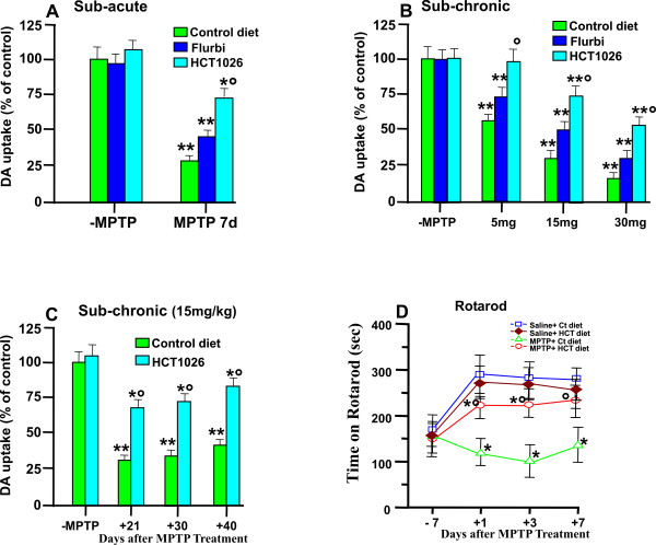
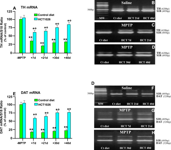
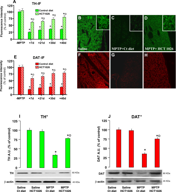
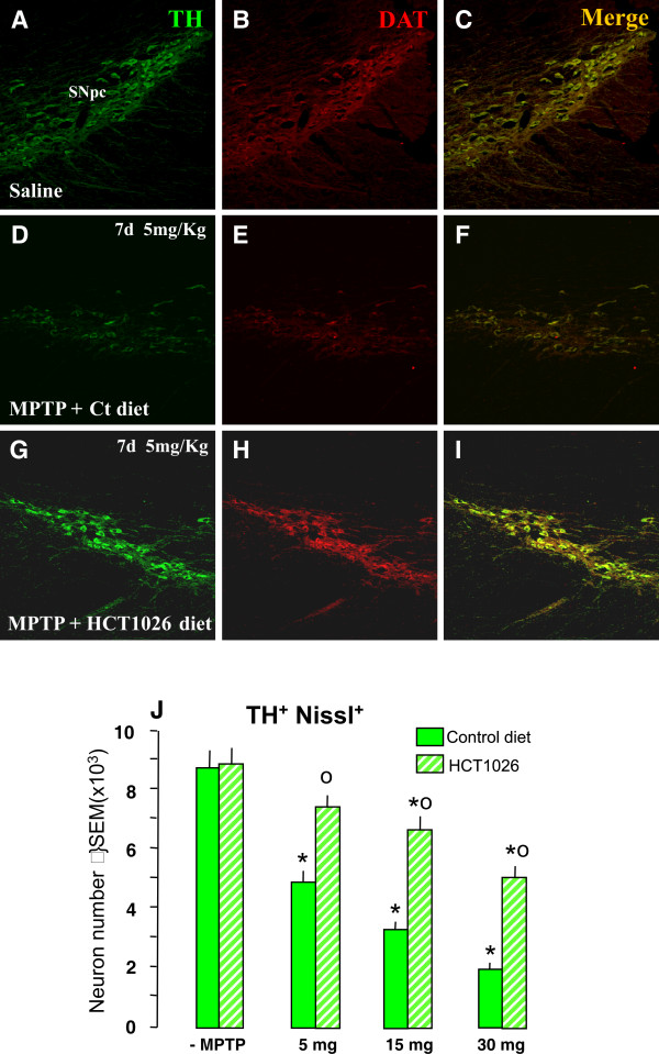
Methods: Ageing mice were fed with a control, flurbiprofen, or HCT1026 diet starting ten days before MPTP administration and continuing for all the experimental period. Striatal high affinity synaptosomal dopamine up-take, motor coordination assessed with the rotarod, tyrosine hydroxylase (TH)- and dopamine transporter (DAT) fiber staining, stereological cell counts, immunoblotting and gene expression analyses were used to assess MPTP-induced nigrostriatal DAergic toxicity and glial activation 1-40 days post-MPTP.

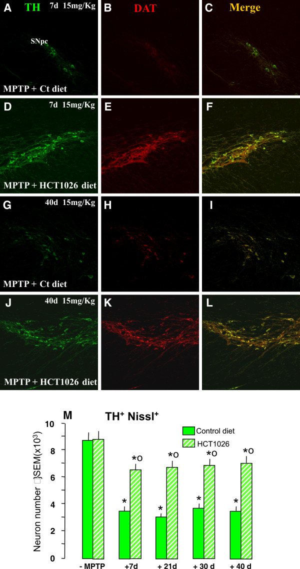
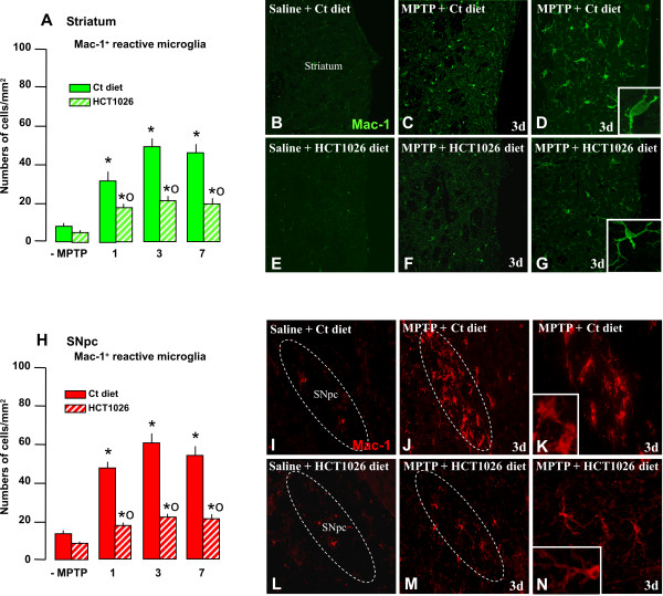
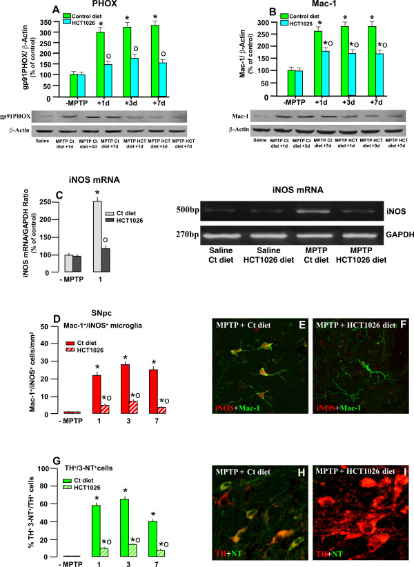
Results: HCT1026 was well tolerated and did not cause any measurable toxic effect, whereas flurbiprofen fed mice showed severe gastrointestinal side-effects. HCT1026 efficiently counteracted motor impairment and reversed MPTP-induced decreased synaptosomal [3H]dopamine uptake, TH- and DAT-stained fibers in striatum and TH+ neuron loss in substantia nigra pars compacta (SNpc), as opposed to age-matched mice fed with a control diet. These effects were associated to a significant decrease in reactive macrophage antigen-1 (Mac-1)-positive microglial cells within the striatum and ventral midbrain, decreased expression of iNOS, Mac-1 and NADPH oxidase (PHOX), and downregulation of 3-Nitrotyrosine, a peroxynitrite finger print, in SNpc DAergic neurons.

Conclusions: Oral treatment with HCT1026 has a safe profile and a significant efficacy in counteracting MPTP-induced dopaminergic (DAergic) neurotoxicity, motor impairment and microglia activation in ageing mice. HCT1026 provides a novel promising approach towards the development of effective pharmacological neuroprotective strategies against PD.


Figures

Figure 1
Figure 1
HCT1026 inhibits MPTP-induced loss of high affinity synaptosomial [3H]DA uptake and reverses motor impairment. Young and ageing C57Bl/6 mice fed with a control, flurbiprofen or HCT1026 diets (30 mg kg-1) starting at -10 d, underwent an MPTP treatment according to the subacute (A) or subchronic (B), injection paradigms, as described. Age-matched mice fed with the different diets received physiologic saline (NaCl, 10 ml kg-1) and served as controls. Seven days after MPTP discontinuance, loss of DAergic functionality was assessed in striatum measuring high affinity synaptosomial striatal [3DA] uptake [8]. HCT1026 prooved to be more potent than its parent compound in counteracting MPTP-induced decreases in striatal DA uptake levels in both the subacute (A) and subchronic (B) protocols. Differences were analyzed by ANOVA followed by Newman-Keuls test, and considered significant when p < 0.05. **p < 0.05 vs saline, ° p < 0.05 vs MPTP + control diet. C. Ageing mice fed with a control or HCT1026 diets, were submitted to the subchronic MPTP regimen, and striatal DA uptake levels measured 21, 30 and 40 d after MPTP (n = 6/time point). Note the long-lasting counteraction of MPTP-induced striatal toxicity in mice fed with HCT1026 as opposed to the control diet. D: Motor performances on Rotarod of saline- and MPTP-treated mice (n = 10/group) fed with a control or HCT1026 diets. Time of permanence on revolving bars (ordinate) are plotted against pre- and post-treatment days (5 trials/day) during which experiments were performed. Mean and SEM values are reported. Establishment of a motor deficit measured 1-7 dpt, is counteracted by HCT1026. Differences were analyzed as above. ** p < 0.05 vs saline; ° p < 0.05 vs MPTP + control diet.
Figure 2
Figure 2
HCT1026 inhibits MPTP-induced loss of striatal TH and DAT mRNAs expression. Ageing (9-11 month-old) C57Bl/6 mice fed with a control (ct) or HCT1026 diets (30 mg kg-1) starting at -10 d, underwent an MPTP treatment according to the subchronic injection paradigm, as described. Age-matched mice fed with the different diets received physiologic saline and served as controls. Mice were sacrificed at different time-intervals after MPTP. Striatal tissue samples were processed for semi-quantitative RT-PCR analysis as described. Total RNA isolated and cDNA synthesized using Retroscript Kit (see Materials and Methods) following the manufacturer's directions. The 250 ng of cDNA were used for PCR, by using Super Taq DNA polymerase with specific primer pairs for TH (620 bp) and DAT (328 bp), and Classic S18 Standard (495 bp). Samples from PCR reactions were separated electrophoretically on 2% agarose gel containing 0,2 μg/ml of ethidium bromide (B-D, F-H). Fluorescent bands of amplified gene products were captured by using Gel Logic 200 Imaging System (Kodak), values normalized against S18 and ratios expressed as percent of control, within each experimental group (A, E). Differences were analyzed by ANOVA followed by Newman-Keuls test, and considered significant when p < 0.05. ** vs saline; ° p < 0.05 vs MPTP + control diet. Note the marked and long-lasting downregulation of TH (A,B,C,D) and DAT (E,F,G,H) mRNA transcript levels in striatal samples from ageing mice submitted to the subchronic MPTP regimen and the significant counteraction afforded by HCT1026.
Figure 3
Figure 3
HCT1026 inhibits MPTP-induced loss of striatal TH- and DAT- proteins by immunohistochemistry and western blotting. Ageing C57Bl/6 mice fed with a control (ct) or HCT1026 diets starting at -10 d, underwent an MPTP treatment, as described. At different time-intervals, mice were anesthetized and rapidly perfused, the brains were carefully removed and processed for immunohistochemistry, as described. TH- (A) and DAT-(E) IR in striatum (Str) assessed by immunofluorescent staining and image analysis by confocal Laser microscopy in ageing mice fed with ct or HCT1026 diets, treated with saline or MPTP (n = 5/time point). Fluorescence intensity values (FI, means ± S.E.M.) are expressed as % of saline. **p < 0.05 vs saline, °p < 0.05 vs MPTP fed with control diet. B-H: Representative confocal images show loss of TH-IF (revealed by FITC, green) in Str of MPTP mice fed with a ct diet at 40 dpt (C) and a substantial rescue of TH- (D) by HCT1026. F-H: Representative confocal images show loss of DAT-IF (revealed by FITC, green) in Str of MPTP mice fed with a ct diet at 40 dpt (G) and a substantial rescue of DAT-IF (H) by HCT1026. E-F: For western blot analysis, at 40 d after saline or MPTP injections in mice fed with the ct or HCT1026 diets, mice were sacrificed and striatal tissue samples processed for WB, as described. The data from experimental bands were normalized to β-actin, before statistical analysis of variance and values expressed as % of saline-injected controls, within each respective group. Note the significant decreased TH (I) and DAT (J) protein levels in MPTP mice fed with a ct diet, whereas a recovery was observed in HCT1026 fed mice. *p < 0.05 vs saline; *° p < 0.05 vs MPTP fed with ct.
Figure 4
Figure 4
HCT1026 preventive administration inhibits MPTP-induced dose-dependent loss of TH+ cell bodies in SNpc. Ageing mice fed with a ct or HCT1026 diets were submitted to the subchronic MPTP (5 mg, 15 mg or 30 mg kg-1, for 5 consecutive d) regimen, and DAergic cell survival studied after 7 d. A-E: Representative confocal images of dual staining with TH- (green) and DAT- (red) -Abs of coronal midbrain sections at the level of the SNpc 7 d after MPTP. Note the significant protection afforded by HCT10926 preventive treatment in mice treated with the 5 mg kg-1 dose (see panels G-I) as compared to MPTP mice fed with a ct diet (D-F). F. Survival of DAergic cell bodies in SNpc. The total number of TH+ and Nissl + neurons was counted throught the entire rostro-caudal axis of the SNpc. Treatment groups were averaged (means ± S.E.M.) * p < 0.05 vs saline; *°p < 0.05, vs MPTP mice fed with a ct diet. HCT1026 significantly reduced the dose-dependent decrease of TH+ and Nissl + neurons.
Figure 5
Figure 5
Long-lasting protection of DAergic cell bodies in HCT1026 fed mice. Ageing mice fed with a ct or HCT1026 diet were submitted to MPTP subchronic regimen (15 mg kg-1 for 5 cosecutive d) and sacrificed at differet time-intervals after MPTP. A-L: Representative confocal images of dual staining with TH- (green) and DAT- (red) -Abs of coronal midbrain sections at the level of the SNpc 7 and 40 d after MPTP. As observed, MPTP mice fed with a ct diet show a marked loss of TH+DAT+ neurons at 7 d (see panels A-C) and fail to recover 40 d following MPTP injury (G-I). By contrast, HCT1026 afforded a significant and lonlasting protection (see panels D-F and J-L). M: The total number of TH+ and Nissl + neurons was counted throught the entire rostro-caudal axis of the SNpc. Treatment groups were averaged (means ± S.E.M.) * p < 0.05 vs saline; *°p < 0.05, vs MPTP mice fed with a ct diet. HCT1026 significantly reduced the decrease of TH+ and Nissl + neurons observed up to 40 d in mice fed with a ct diet.
Figure 6
Figure 6
HCT1026 inhibits MPTP-induced increased reactive Mac-1+ microglia in striatum and SNpc. Ageing (9-11 month-old) C57Bl/6 mice fed with a control (ct) or HCT1026 diets (30 mg kg-1) starting at -10 d, underwent an MPTP treatment, as described. At different time-intervals mice were anesthetized and rapidly perfused, the brains processed for immunohistochemistry. Coronal sections at the level of the striatum and SNpc were stained with Mac-1-Ab to localize microglial cells. A-B: Reactive (ameboid-like) microglial cells were counted at different time-intervals after saline and MPTP injection (n = 4/experimental group) in mice fed with a ct or HCT1026 diets, within both Str (A-G) and SNpc (H-N). Differences were analyzed by ANOVA followed by Newman-Keuls test, and considered significant when p < 0.05. **p < 0.05 vs saline; *° p < 0.05 vs MPTP mice fed with ct diet. B-E: Representative confocal images of Mac-1 staining (green) in saline (B), 3 d after MPTP in mice fed with ct diet (C, 20× and D, 40×) or HCT1026 (F, 20× and G, 40×). Insets (100×) show microglia morphologic appearance. I-N: Representative confocal images of Mac-1 staining (red) in SNpc. Note the high density of reactive Mac-1+ cells with rounded cell bodies and short, thick processes 3 d after MPTP administration in mice fed with a ct diet (J-K), as compared to Mac-1 microglia of mice fed with HCT1026 (M-N), exhibiting a more elongated cell body and long ramified processes.
Figure 7
Figure 7
HCT1026 inhibits MPTP-induced increased Mac-1, PHOX, iNOX and 3-Nitrotyrosine expression in SNpc. A-B: Western blotting of phagocyte oxidase PHOX (A) and Mac-1 (B) within the VM at different time-intervals after saline or MPTP injection in mice fed with the control or HCT1026 diet. The data from experimental bands were normalized to β-actin, before statistical analysis of variance. Values are expressed as % of saline-injected controls. Differences were analyzed by ANOVA followed by Newman-Keuls test, and considered significant when p < 0.05. * p < 0.05 compared to saline; ° p < 0.05 vs MPTP fed with the control diet. C. Semi-quantitative RT-PCR for iNOS. The 250 ng of cDNA were used for PCR, by using Super Taq DNA polymerase with specific primer pairs for iNOS (500 bp) and Classic GADPH Standard (270 bp). Samples from PCR reactions were processed as described. Fluorescent bands of amplified gene products were analyzed, the values normalized against GADPH and ratios expressed as % of control, within each experimental group (C), see text for details. Differences were analyzed by ANOVA as above. ** p < 0.05 vs saline; ° p < 0.05 vs MPTP + control diet. D: Mean numbers of Mac1+iNOS+ cells within the SNpc in saline and MPTP mice fed with the control or HCT1026 diet. Cell counts obtained as described in Material section. E-F: Representative confocal images showing double staining with Mac-1 (green) and iNOS (red) in MPTP mice fed with the control (E) or HCT1026 (F) diets. G: Percent (%) of TH+ neurons colocalizing with 3-nitrotyrosine (3-NT), a peroxynitrite footprint. Dual stained TH+ 3-NT+ neurons were counted as described and values expressed as % of total TH+ neurons. H-I: Representative confocal images showing dual immunostaining with 3-NT (green) and TH (red) in MPTP mice fed with a ct diet (H) showing that a large proportion of DAergic neurons colocalize (orange-to-yellow) as opposed to TH neurons of mice fed with HCT1026 (E) where no colocalization was observed in the large part of SNpc neurons.

Similar articles

Cited by

References

    1. Tedroff JM. Functional consequences of dopaminergic degeneration in Parkinson's disease. Adv Neurol. 1999;80:67–70. - PubMed
    1. Rascol O, Payoux P, Ory F, Ferriera JJ, Brefel-Courbon C, Montastruc JL. Limitations of current Parkinson's disease therapy. Ann Neurol. 2003;53(Suppl 3):S3–12. doi: 10.1002/ana.10513. - DOI - PubMed
    1. Warner TT, Schapira AHV. Genetic and environmental factors in the cause of Parkinson's disease. Ann Neurol. 2003;53:S16–S25. doi: 10.1002/ana.10487. - DOI - PubMed
    1. Hindle JV. Ageing, neurodegeneration and Parkinson's disease. Age Ageing. 2010;39:156–161. doi: 10.1093/ageing/afp223. - DOI - PubMed
    1. McGeer P, McGeer E. Inflammation and neurodegeneration in Parkinson's disease. Parkinsonism Relat Disord. 2004;10:S3–7. doi: 10.1016/j.parkreldis.2004.01.005. - DOI - PubMed

MeSH terms