. 2010 Nov 23:9:301.
doi: 10.1186/1476-4598-9-301.

Melanoma-associated Chondroitin Sulfate Proteoglycan (MCSP)-targeted delivery of soluble TRAIL potently inhibits melanoma outgrowth in vitro and in vivo

Affiliations

Melanoma-associated Chondroitin Sulfate Proteoglycan (MCSP)-targeted delivery of soluble TRAIL potently inhibits melanoma outgrowth in vitro and in vivo

Marco de Bruyn et al. Mol Cancer. .

Abstract

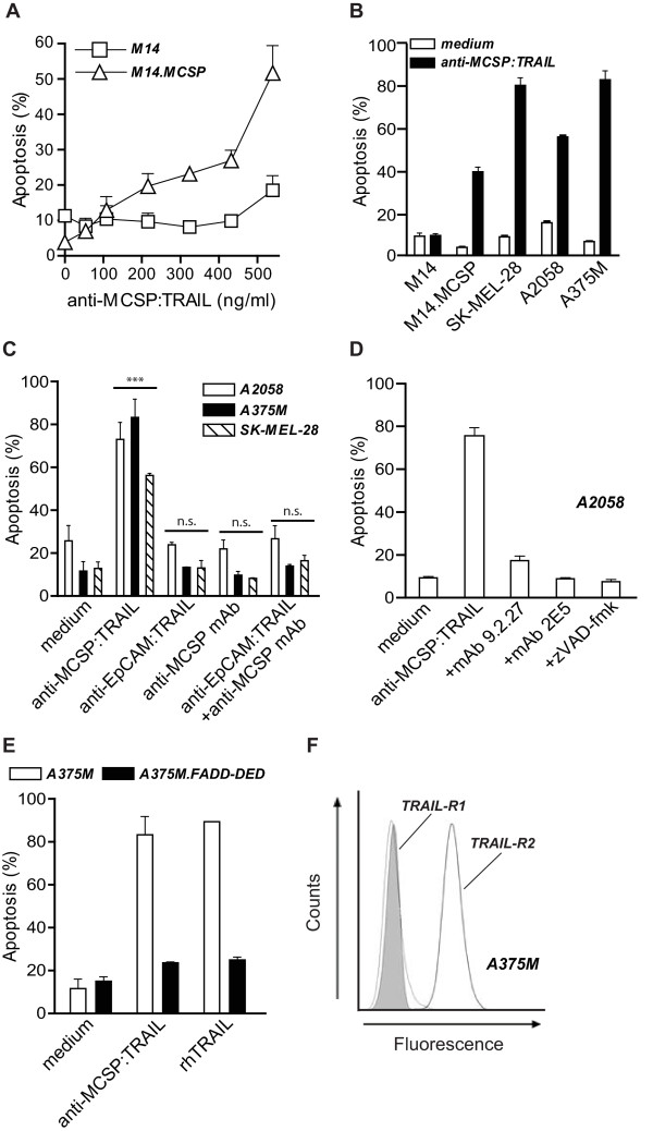
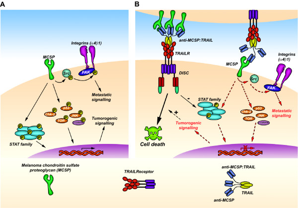
Background: Advanced melanoma is characterized by a pronounced resistance to therapy leading to a limited patient survival of ~6 - 9 months. Here, we report on a novel bifunctional therapeutic fusion protein, designated anti-MCSP:TRAIL, that is comprised of a melanoma-associated chondroitin sulfate proteoglycan (MCSP)-specific antibody fragment (scFv) fused to soluble human TRAIL. MCSP is a well-established target for melanoma immunotherapy and has recently been shown to provide important tumorigenic signals to melanoma cells. TRAIL is a highly promising tumoricidal cytokine with no or minimal toxicity towards normal cells. Anti-MCSP:TRAIL was designed to 1. selectively accrete at the cell surface of MCSP-positive melanoma cells and inhibit MCSP tumorigenic signaling and 2. activate apoptotic TRAIL-signaling.

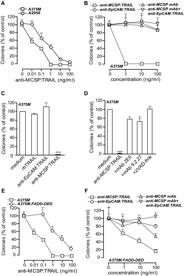
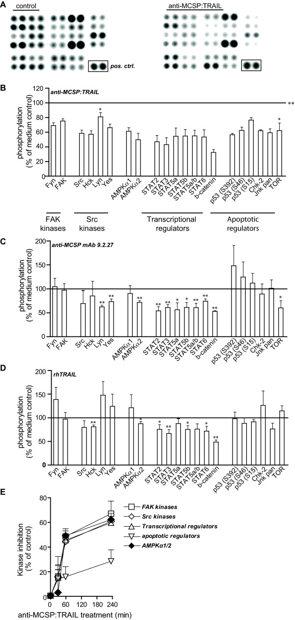
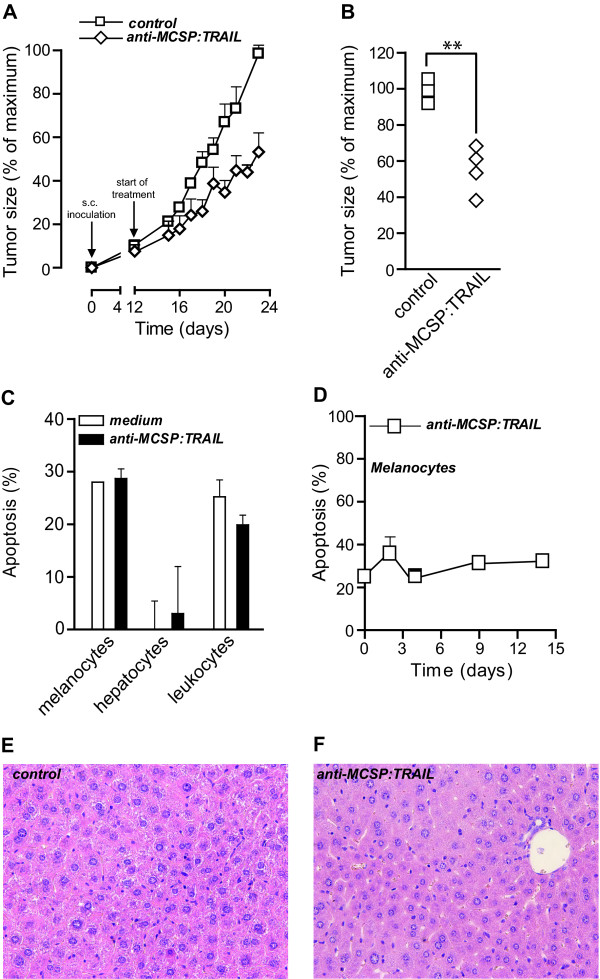
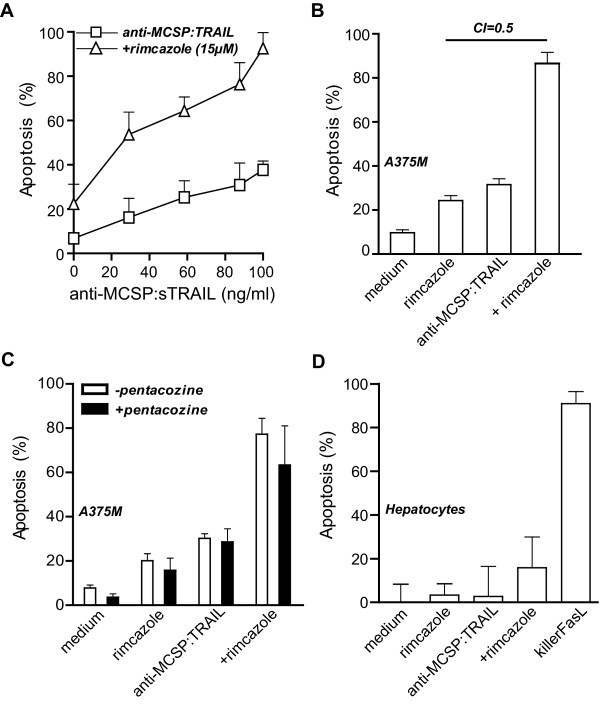
Results: Treatment of a panel of MCSP-positive melanoma cell lines with anti-MCSP:TRAIL induced TRAIL-mediated apoptotic cell death within 16 h. Of note, treatment with anti-MCSP:sTRAIL was also characterized by a rapid dephosphorylation of key proteins, such as FAK, implicated in MCSP-mediated malignant behavior. Importantly, anti-MCSP:TRAIL treatment already inhibited anchorage-independent growth by 50% at low picomolar concentrations, whereas > 100 fold higher concentrations of non-targeted TRAIL failed to reduce colony formation. Daily i.v. treatment with a low dose of anti-MCSP:TRAIL (0.14 mg/kg) resulted in a significant growth retardation of established A375 M xenografts. Anti-MCSP:TRAIL activity was further synergized by co-treatment with rimcazole, a σ-ligand currently in clinical trials for the treatment of various cancers.

Conclusions: Anti-MCSP:TRAIL has promising pre-clinical anti-melanoma activity that appears to result from combined inhibition of tumorigenic MCSP-signaling and concordant activation of TRAIL-apoptotic signaling. Anti-MCSP:TRAIL alone, or in combination with rimcazole, may be of potential value for the treatment of malignant melanoma.


Figures

Figure 1
Figure 1
MCSP-restricted induction of apoptosis by anti-MCSP:TRAIL. A M14 and M14.MCSP cells were treated with increasing concentrations of anti-MCSP:TRAIL for 16 h and apoptosis was assessed by ∆ψ. B MCSP-negative cell line M14 and MCSP-positive cell lines M14.MCSP, SK-MEL-28, A2058 and A375M were treated with 500 ng/ml anti-MCSP:TRAIL for 16 h and apoptosis was assessed by ∆ψ. C A375M, A2058 and SK-MEL-28 cells were treated with equimolar concentrations of anti-MCSP:TRAIL, anti-EpCAM:TRAIL, anti-MCSP mAb or anti-EpCAM:TRAIL+anti-MCSP mAb for 16 h and apoptosis was assessed by ∆ψ. D A2058 cells were treated with 500 ng/mL anti-MCSP:TRAIL in the absence or presence of parental MCSP-blocking mAb 9.2.27, TRAIL-neutralizing mAb 2E5 or pan-caspase inhibitor zVAD-fmk for 16 h and apoptosis was assessed by ∆ψ. E A375M and A375M.FADD-DED cells were treated with anti-MCSP:TRAIL or rhTRAIL for 16 h and apoptosis was assessed by ∆ψ. F A375M cells were incubated with anti-TRAIL-R1 (thin line) or anti-TRAIL-R2 (thick line) mAb and expression of TRAIL-R1 and TRAIL-R2 was analyzed by flow cytometry. Shaded area indicates the fluorescence signal when cells were incubated with fluorescent-labeled secondary antibody alone.
Figure 2
Figure 2
Inhibition of anchorage-independent growth by anti-MCSP:TRAIL. A A375M and A2058 cells were treated with increasing concentrations of anti-MCSP:TRAIL for 21 days and colony formation was assessed. B A375M cells were treated with increasing concentrations of anti-MCSP:TRAIL, anti-EpCAM:TRAIL, anti-MCSP mAb or anti-EpCAM:TRAIL+anti-MCSP mAb for 21 days and colony formation was assessed. C A375M cells were treated with rhTRAIL, anti-EpCAM:TRAIL or anti-MCSP:TRAIL for 21 days and colony formation was assessed. D A375M cells were treated with 100 ng/ml anti-MCSP:TRAIL in the absence or presence of TRAIL-neutralizing mAb 2E5, parental anti-MCSP mAb 9.2.27 or pan-caspase inhibitor zVAD-fmk for 21 days and colony formation was assessed. E A375M and A375M.FADD-DED cells were treated with increasing concentrations of anti-MCSP:TRAIL for 21 days and colony formation was assessed. F A375M.FADD-DED cells were treated with increasing concentrations of anti-MCSP:TRAIL, anti-EpCAM:TRAIL, anti-MCSP mAb or anti-EpCAM:TRAIL+anti-MCSP mAb for 21 days and colony formation was assessed.
Figure 3
Figure 3
Anti-MCSP:TRAIL triggers dephosphorylation of cellular proteins. A375M cells were treated with anti-MCSP:TRAIL, anti-MCSP mAb 9.2.27, rhTRAIL or left untreated for 1 h. Whole cell lysates were analyzed for phosphorylation of 48 cellular kinases. A Phosphorylation of 28 kinases in untreated cells (left panel) or anti-MCSP:TRAIL-treated cells (right panel) are depicted. Images are representative of three independent experiments. B, C, D Changes in the phosphorylation of 21 kinases after treatment with B anti-MCSP:TRAIL, C anti-MCSP mAb 9.2.27 or D rhTRAIL are expressed as a percentage of the phosphorylation measured in untreated cells. E A375M cells were treated with anti-MCSP:TRAIL for 0, 30, 60 or 240 min. Whole cell lysates were analyzed for phosphorylation of 48 cellular kinases. Changes in phosphorylation of the different kinase groups as indicated in panel B are depicted. *, p < 0.05; **, p < 0.001; ***, p < 0.0001.
Figure 4
Figure 4
Anti-tumor activity of anti-MCSP:TRAIL towards A375M xenografts. A Mice were inoculated with A375M tumor cells at day 0 and developed xenografts of ~ 50 mm3 after 12 days. Starting at day 12, mice were injected daily with saline (n = 4) or anti-MCSP:TRAIL (0.14 mg/kg, n = 4) and tumor size was measured daily using electronic caliper measurements. B Tumor size at day 23. C MCSP-positive melanocytes and MCSP-negative leukocytes and hepatocytes were treated with 500 ng/ml anti-MCSP:TRAIL for 16 h and apoptosis was assessed by ∆ψ. D Melanocytes were treated with 4 μg/mL anti-MCSP:TRAIL for up to 14 days and apoptosis was assessed by ∆ψ at time points indicated. E Liver pathology of mice carrying A375M xenografts was examined for Sham-treated mice or F anti-MCSP:TRAIL-treated mice.
Figure 5
Figure 5
Synergistic induction of apoptosis by anti-MCSP:TRAIL and rimcazole A. A375M cells were treated with increasing concentrations of anti-MCSP:TRAIL in the presence or absence of rimcazole for 16 h and apoptosis was assessed by ∆ψ. B A375M cells were treated with rimcazole (15 μM), anti-MCSP:TRAIL (100 ng/ml) or both for 16 h, apoptosis was assessed by ∆ψ and the cooperativity index (CI) was calculated as indicated in the materials and methods section. C A375M cells were treated with rimcazole (15 μM), anti-MCSP:TRAIL (100 ng/mL) or both in the presence or absence of pentacozine (200 nM) for 16 h and apoptosis was assessed by ∆ψ. D Hepatocytes were treated with, rimcazole (15 μM), anti-MCSP:TRAIL (100 ng/ml) or both for 16 h and apoptosis was assessed by ∆ψ. KillerFasL (100 ng/ml)was used as a positive control for hepatocyte toxicity.
Figure 6
Figure 6
Schematic representation of the anti-melanoma activity of anti-MCSP:TRAIL. A Direct or indirect MCSP-mediated signaling involves (integrin-dependent) FAK and Src phosphorylation and maintains phosphorylation of downstream transcription factors and apoptotic modulators. MCSP thus contributes to pro-survival and metastatic signals. B Binding of anti-MCSP:TRAIL inhibits MCSP-mediated signaling and concomitant activation of pro-metastatic/-survival signals and thereby sensitizes cells to apoptosis induction via binding of the TRAIL moiety to TRAILR.

Similar articles

Cited by

References

    1. Balch C, Buzaid A, Soong S, Atkins M, Cascinelli N, Coit D, Fleming I, Gershenwald J, Houghton A, Kirkwood J. et al.Final Version of the American Joint Committee on Cancer Staging System for Cutaneous Melanoma. J Clin Oncol. 2001;19:3635–3648. - PubMed
    1. Helmbach H, Sinha P, Schadendorf D. Human melanoma: drug resistance. Recent Results Cancer Res. 2003;161:93–110. - PubMed
    1. Campoli MR, Chang CC, Kageshita T, Wang X, McCarthy JB, Ferrone S. Human high molecular weight-melanoma-associated antigen (HMW-MAA): a melanoma cell surface chondroitin sulfate proteoglycan (MSCP) with biological and clinical significance 1. Crit Rev Immunol. 2004;24:267–296. doi: 10.1615/CritRevImmunol.v24.i4.40. - DOI - PubMed
    1. Vergilis I, Szarek M, Ferrone S, Reynolds S. Presence and Prognostic Significance of Melanoma-Associated Antigens CYT-MAA and HMW-MAA in Serum of Patients with Melanoma. J Investig Dermatol. 2005;125:526–531. doi: 10.1111/j.0022-202X.2005.23798.x. - DOI - PubMed
    1. Eisenmann KM, McCarthy JB, Simpson MA, Keely PJ, Guan JL, Tachibana K, Lim L, Manser E, Furcht LT, Iida J. Melanoma chondroitin sulphate proteoglycan regulates cell spreading through Cdc42, Ack-1 and p130cas. Nat Cell Biol. 1999;1:507–513. doi: 10.1038/70302. - DOI - PubMed

Publication types

MeSH terms

Substances