. 2010 Nov 23:3:113.
doi: 10.1186/1756-3305-3-113.

Bovipain-2, the falcipain-2 ortholog, is expressed in intraerythrocytic stages of the tick-transmitted hemoparasite Babesia bovis

Affiliations

Bovipain-2, the falcipain-2 ortholog, is expressed in intraerythrocytic stages of the tick-transmitted hemoparasite Babesia bovis

María Mesplet et al. Parasit Vectors. .

Abstract

Background: Cysteine proteases have been shown to be highly relevant for Apicomplexan parasites. In the case of Babesia bovis, a tick-transmitted hemoparasite of cattle, inhibitors of these enzymes were shown to hamper intraerythrocytic replication of the parasite, underscoring their importance for survival.

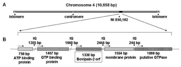
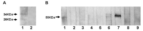
Results: Four papain-like cysteine proteases were found to be encoded by the B. bovis genome using the MEROPS database. One of them, the ortholog of Plasmodium falciparum falcipain-2, here named bovipain-2, was further characterized. Bovipain-2 is encoded in B. bovis chromosome 4 by an ORF of 1.3 kb, has a predicted molecular weight of 42 kDa, and is hydrophilic with the exception of a transmembrane region. It has orthologs in several other apicomplexans, and its predicted amino acid sequence shows a high degree of conservation among several B. bovis isolates from North and South America. Synteny studies demonstrated that the bovipain-2 gene has expanded in the genomes of two related piroplasmids, Theileria parva and T. annulata, into families of 6 and 7 clustered genes respectively. The bovipain-2 gene is transcribed in in vitro cultured intra-erythrocyte forms of a virulent and an attenuated B. bovis strain from Argentina, and has no introns, as shown by RT-PCR followed by sequencing. Antibodies against a recombinant form of bovipain-2 recognized two parasite protein bands of 34 and 26 kDa, which coincide with the predicted sizes of the pro-peptidase and mature peptidase, respectively. Immunofluorescence studies showed an intracellular localization of bovipain-2 in the middle-rear region of in vitro cultured merozoites, as well as diffused in the cytoplasm of infected erythrocytes. Anti-bovipain-2 antibodies also reacted with B. bigemina-infected erythrocytes giving a similar pattern, which suggests cross-reactivity among these species. Antibodies in sera of two out of six B. bovis-experimentally infected bovines tested, reacted specifically with recombinant bovipain-2 in immunoblots, thus demonstrating expression and immunogenicity during bovine-infecting stages.

Conclusions: Overall, we present the characterization of bovipain-2 and demonstrate its in vitro and in vivo expression in virulent and attenuated strains. Given the involvement of apicomplexan cysteine proteases in essential parasite functions, bovipain-2 constitutes a new vaccine candidate and potential drug target for bovine babesiosis.


Figures

Figure 1
Figure 1
Localization of the bovipain-2 gene in the genome of the Babesia bovis strain T2Bo. A. Schematic representation of chromosome 4 of the B. bovis strain T2Bo showing the relative localization of the bovipain-2 encoding gene. The localization of the centromere and telomeres are indicated by arrows. B. Organization of the B. bovis ~39.4 kbp genomic region containing the locus of bovipain-2. The orientation of the open reading frames (ORF) is shown with black arrows. IG: intergenic regions.
Figure 2
Figure 2
Domain prediction of bovipain-2. Putative functional domains in bovipain-2 were predicted based on the amino acid sequence of the T2B strain. The numbers indicate the eukaryotic thiol (cysteine) proteases cysteine (1); histidine (2) and asparagine (3) catalytic regions. Amino acids in bold represent active sites (Q254, C260, H389, N411). Sequence alignments of these three regions in several Babesia, Theileria and Plasmodium spp. are shown. Arrows indicate amino acids of the active site.
Figure 3
Figure 3
Phylogenetic and synteny relationships between B. bovis, T. annulata and T. parva C1 cysteine peptidases. A. Phylogenetic relationships of cysteine peptidases of B. bovis and their Theileria annulata and T. parva orthologs/paralogs as analyzed by neighbor joining. The tree is drawn to scale, with branch lengths in the same units as those of the evolutionary distances used to infer the phylogenetic tree. The evolutionary distances were computed using the Poisson correction method. Deletion of all positions containing gaps and missing data resulted in a total of 171 positions in the final dataset. The percentage of replicate trees in which the associated taxa clustered together in the bootstrap test (1000 replicates) is shown next to the branches. The GenBank reference sequence number of each protein is shown. Bb: B. bovis; Ta: Theileria annulata; Tp: T. parva; Lm: Leishmania major cathepsin L-like protein is used as outgroup. B. Synteny studies of the bovipain-2 encoding genome region with the ortholog/paralogs encoding regions of T. annulata and T. parva. Lines that connect the catalytic region of the bovipain-2 [GenBank: XP_001610695] encoding gene and the catalytic regions of related cysteine proteinases encoding genes of T. annulata, and T. parva represent high scoring sequence pairs (HSPs). Reference protein sequence numbers from right to left: T. annulata, [GenBank: XP_954970, XP_954971, XP_954972, XP_954973, XP_954974, XP_954975, XP_954976], and T. parva, [GenBank: XP_763298, XP_763298, XP_763298, XP_763298, XP_763298, XP_763298]. A small ORF encoding a hypothetical protein [GenBank XP_001610696] in B. bovis has corresponding HSPs in T. annulata and T. parva. In T. annulata this ORF has not been annotated.
Figure 4
Figure 4
Transcription of the bovipain-2 gene. Bovipain-2 transcripts were detected by total RNA extraction of in vitro cultured B. bovis merozoites (BboS2P strain), followed by incubation with reverse transcriptase and PCR amplification of the complete ORF (RT+). To rule out DNA contamination, an equal amount of RNA was not incubated with reverse transcriptase and then treated as above (RT-). 1 kb: DNA size standard. The size of the obtained amplicon is marked at the left.
Figure 5
Figure 5
Expression and immunogenicity of bovipain-2. A. Expression of bovipain-2 in in vitro cultured merozoites. Aliquots of a merozoite suspension of the BboS2P strain of B. bovis were electrophoresed, blotted and exposed to antibodies against (1) recombinant bovipain-2, or (2) normal serum. Antibody binding was detected by incubation with peroxidase-labeled anti-mouse IgG and chemiluminiscence. Bands of 34 and 26 kDa correspond to the expected sizes of bovipain-2 pro-protein and mature protein, respectively. B. Immunogenicity of bovipain-2 in B. bovis-experimentally infected cattle. Stripes with blotted, partially purified bovipain-2 were incubated with sera from different bovines experimentally infected (day 63 p.i.) with the BboR1A strain (stripes 1-3) or the BboS2P strain of B. bovis (4-6); non infected bovine sera (8-9); or a monoclonal antibody that reacts with the histidine tag of the recombinant protein (7). A band of 50-55 kDa was recognized in samples 1, 6 and 7 by reaction with peroxidase-labeled species-specific secondary antibodies, followed by chemiluminiscence detection.
Figure 6
Figure 6
Localization of bovipain-2 in merozoites. Erythrocytes infected with B. bovis strain BboS2P (A, D, E); B. bovis, strain RAD (B); B. bigemina, Mexico strain (C), or non infected erythrocytes (F) were incubated with murine anti-bovipain-2 antibodies (A, B, C, F); anti-B. bovis RAP-1 monoclonal antibody 23/70.174 (D) or pre-immune murine serum (E); followed by detection with FITC- (A,D,E,F) or Alexa Fluor 488- (B,C) labeled anti-murine IgG, and observation by epifluorescene, with 400 × (A,D,E,F) or 1000 × magnification (B,C). Bovipain-2-associated fluorescence was observed as a round spot inside merozoites and within the erythrocyte cytoplasm (A,B,C), different from the punctuated fluorescence associated to RAP-1 (D). No cross-reactivity with erythrocyte proteins was detected (F); and no unspecific reaction was observed with pre-immune mouse serum (E).

Similar articles

Cited by

References

    1. Bock R, Jackson L, de Vos A, Jorgensen W. Babesiosis of cattle. Parasitology. 2004;129(Supp l):S247–269. doi: 10.1017/S0031182004005190. - DOI - PubMed
    1. Florin-Christensen M, Schnittger L, Dominguez M, Mesplet M, Rodriguez A, Ferreri L, Asenzo G, Wilkowsky S, Farber M, Echaide I, Suarez C. Search for Babesia bovis vaccine candidates. Parassitologia. 2007;49(Suppl 1):9–12. - PubMed
    1. Brayton KA, Lau AO, Herndon DR, Hannick L, Kappmeyer LS, Berens SJ, Bidwell SL, Brown WC, Crabtree J, Fadrosh D, Feldblum T, Forberger HA, Haas BJ, Howell JM, Khouri H, Koo H, Mann DJ, Norimine J, Paulsen IT, Radune D, Ren Q, Smith RK Jr, Suarez CE, White O, Wortman JR, Knowles DP Jr, McElwain TF, Nene VM. Genome sequence of Babesia bovis and comparative analysis of apicomplexan hemoprotozoa. PLoS Pathog. 2007;3:1401–13. doi: 10.1371/journal.ppat.0030148. - DOI - PMC - PubMed
    1. Krause PJ, Daily J, Telford SR, Vannier E, Lantos P, Spielman A. Shared features in the pathobiology of babesiosis and malaria. Trends Parasitol. 2007;23:605–610. doi: 10.1016/j.pt.2007.09.005. - DOI - PubMed
    1. Sajid M, McKerrow JH. Cysteine proteases of parasitic organisms. Mol Biochem Parasitol. 2002;120:1–21. doi: 10.1016/S0166-6851(01)00438-8. - DOI - PubMed

LinkOut - more resources