Comparative Study
. 2010 Nov 18;68(4):682-94.
doi: 10.1016/j.neuron.2010.09.030.

The transcription factor Pax6 regulates survival of dopaminergic olfactory bulb neurons via crystallin αA

Affiliations
Comparative Study

The transcription factor Pax6 regulates survival of dopaminergic olfactory bulb neurons via crystallin αA

Jovica Ninkovic et al. Neuron. .

Abstract

Most neurons in the adult mammalian brain survive for the entire life of an individual. However, it is not known which transcriptional pathways regulate this survival in a healthy brain. Here, we identify a pathway regulating neuronal survival in a highly subtype-specific manner. We show that the transcription factor Pax6 expressed in dopaminergic neurons of the olfactory bulb regulates the survival of these neurons by directly controlling the expression of crystallin αA (CryαA), which blocks apoptosis by inhibition of procaspase-3 activation. Re-expression of CryαA fully rescues survival of Pax6-deficient dopaminergic interneurons in vivo and knockdown of CryαA by shRNA in wild-type mice reduces the number of dopaminergic OB interneurons. Strikingly, Pax6 utilizes different DNA-binding domains for its well-known role in fate specification and this role of regulating the survival of specific neuronal subtypes in the mature, healthy brain.


Figures

Fig. 1
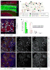
Fig. 1. Pax6 function is necessary for the survival of dopaminergic periglomerular neurons
(A–B) Schematic representation of the main neuronal layers in the OB (A) and neuronal subtypes in the GL (B, region depicted as white square in A). (C) Fluorescence micrograph depicting that virtually all TH-immunoreactive dopaminergic periglomerular neurons (red) also contain Pax6 (green) in 3 month old mice. (D–F) Loss of Pax6 in homozygous Pax6fl/fl mice after DAT::Cre mediated recombination and indicated by GFP reporter (green) results in reduced reporter+ (D, E′,F′) and TH+ (red) (D,E″,F″) cells (11 animals for control and 7 for Pax6fl/fl). (E–F) Fluorescence micrographs depicting the GL (outlined with dotted lines) of Pax6 deficient animals (F) and heterozygote siblings (E) immunostained for TH (red) and GFP reporter (green). Note the reduced number of cells positive for the GFP reporter (indicating Pax6-deficient cells) and for TH in Pax6fl/fl animal (arrows in F). Abbreviations: GL-glomerular layer; EPL-external plexiform layer; ML-mitral cells; GCL-granular cell layer; RMS-rostral migratory stream; IPL-internal plexiform layer; Gm- glomerulus, TH-tyrosine hydroxylase, CR-calretinin, CB-calbindin. Scale bars: 50 μm. ***- p<0.001 and brackets in D represent SEM.
Figure 2
Figure 2. Propidium iodide (PI)/annexinV based cell death analysis of OB cells after DAT::Cre mediated Pax6 deletion
This method is based on the difference in integrity of the plasma membrane in apoptosis and necrosis. One of the earliest features of apoptotic cells is the translocation of the phospholipid phosphatidylserine (PS) from the inner to the outer leaflet of the plasma membrane. Therefore, early apoptotic cells can be efficiently labelled with annexinV, which has a high affinity to PS. Loss of plasma membrane integrity, characterising both necrosis and the late phase of apoptosis, allows passage of propidium iodide (PI) through the plasma membrane and labelling of DNA. Assessing recombined, reporter positive cells (P2 population in C) in the DAT::Cre//GFP//Pax6fl/fl (E) and DAT::Cre//GFP//Pax6fl/+ control animals (D) for PI and annexinV, allowed us to distinguish between live (annexinV-negative, PI-negative), early apoptotic (annexinV-positive, PI-negative) and necrotic/late apoptotic (annexinV-positive, PI-positive) cells (B). (A–B) Dot blots illustrating gate definition for the GFP (A), PI and AnnexinV (B) based on analysis of WT (GFP-negative) not stained cells. (CE) Dot blots showing the analyses of recombined cells (P2 population) in Pax6 mutant (DAT::Cre//GFP//Pax6fl/fl) (E) and its control sibling (D). (F) Histogram depicting the proportion of recombined cells immunoreactive to activated caspase 3 before (1 month) and after (3 month) the loss of Pax6 protein (3 animals analyzed per genotype, per time point and at least 150 cells analyzed per animal). Data are showen as mean value ± SEM.
Fig. 3
Fig. 3. CryαA is sufficient to rescue the survival of Pax6-ablated periglomerular neurons
(A, A′) Micrographs depicting CryαA mRNA expression in the adult brain confined to the OB. (B, C) Micrographs depicting the reduced proportion of Cre+ (red) cells double-labelled for CryαA (green; yellow arrows indicate double+ cells) after loss of Pax6 in DAT::Cre//GFP//Pax6fl/fl mice (C, 20±4% double+; 9 animals) compared to the control (B, 97±3% double+, 6 animals). (D– E) Micrographs depicting dopaminergic neuronal population in the olfactory bulb (TH, red) after lentivirus mediated CryαA knock-down (E, E′) and over-expression of the control shRNA (D, D′). Transduced cells are GFP-immunoreactive and D′ and E′ are magnifications of boxed areas in D and E respectively. (F) Histograms showing the distribution of CryαA (grey bars) or control shRNA (open bars) transduced cells in the OB 8 weeks after transduction (left histogram) and the proportion of dopaminergic neurons (right histogram). Note significant decrease (p < 0.005) in CryαA-deficient cells with dopaminergic identity (2 ± 0.5 % of all transduced cells in the GL, 4 animals analyzed) compared with the control shRNA transduced cells (8 ± 2 % of all transduced cells in the GL, 4 animals analyzed) (G–J) Retrovirus mediated CryαA expression (red in H, I) rescues the loss of dopaminergic PGNs (green as GFP+ by DAT::Cre//GFP, yellow arrow) in DAT::Cre//GFP//Pax6fl/fl mice (3 animals), while control virus expressing DsRed only (G, I) fails to rescue (3 animals). (J) Histogram depicting the AnnexinV-positive, dying cells 6 weeks after the re-expression of CryαA (GFP and dsRed) or control vector (dsRed only) in the Pax6-depleted periglomerular neurons identified by the GFP immunoreactivity (3 animals analyzed). Abbreviations: as in Fig. 1 and Ctx-cortex; LV-lateral ventricle; SEZ-subependymal zone; OB-olfactory bulb. Scale bars: 20 μm in B and C; 50 μm in D, E,G and H and 100 μm in D′ and E′. ***- p< 0.001 and brackets in F and I are SEM.
Fig. 4
Fig. 4. CryαA knock-down induces apoptosis via caspase-3 activation
(A–C) Confocal images showing reduction in numbers of P19 cells transfected with the virus expressing GFP and CryαA-specific shRNA5 (B, B′) compared to the controls (A, A, C, C′). (D) Caspase specific inhibitor ZVAD-fmk blocks the effect of CryαA-specific shRNA5 on number and morphology of P19 cells (compare D and B). (E) Histogram depicting the increase in the proportion of early apoptotic and late apoptotic/necrotic cells after CryαA knock-down as detected by annexinV and PI staining and FACS analyses (3 experiments, 10 000 events per condition). (F) Western blot depicting the activation of procaspase-3 (upper band, 30 kDa) and release of active caspase-3 form (lower bands, 13 and 17 kDa) only after the transient transfection of P19 cells with CryαA specific shRNA5. Abbreviations: hpt-hours after transfection, ***-p<0.001; ****-p< 0.0001. Brackets in E are SEM. Scale bars: 50 μm in A, B, C and d; 10 μm in A′, B′ and C′.
Fig. 5
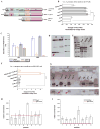
Fig. 5. Pax6 homeodomain activates CryαA expression
(A) Scheme depicting different forms of the Pax6 transcription factor (B). The canonical, 5a and PDless Pax6 forms activate reporter gene expression driven by the 2.1 kb CryαA promoter (Yang et al., 2006), while introduction of a point mutation interfering with the homeodomain DNA-binding in the PDless form (blue ring in A) abolishes the reporter activation (5 experiments) in the Pax6-expressing P19 cell line. (C) The relative abundance of different Pax6 forms measured at RNA (qRT-PCR) and protein (western blot) level in the adult OB and SEZ. (D) Western blot for Pax6 depicting enrichment of PDless Pax6 form (arrow) in the OB compared to the SEZ lysates. Note that both SEZ and OB contain canonical Pax6 and Pax65a (50 – 55 kD bands). (E) HEK293T cells do not express any Pax6 forms. Western blot for Pax6 in non-transfected (right line) and HEK293T cells transfected with constructs expressing Pax6 (left line) or Pax65a (middle line) using AB2237 antibody that recognize all Pax6 forms because of C-terminal binding. (F) In HEK293T cells lacking endogenous Pax6 co-expression of canonical and PDless form of Pax6 is necessary to activate 2.1 kb CryαA promoter (Yang et al., 2006) (red bar), while Pax6, Pax65a and PDless alone do not activate the reporter construct. Note that the point mutation affecting DNA binding capacity of HD abolished CryαA promoter activation. (G) PDless interacts with canonical Pax6 in the OB. Western blot for all Pax6 forms (rabbit anti-Pax6 antibody (see Suppl. Fig. S4)) on canonical Pax6-precipitated with the N-terminal Pax6 antibody (Hybridoma anti-Pax6 antibody (see Suppl. Fig. S4)) in total lysates of the adult OB. No signal for Pax6 forms was detected in the wash fraction or immunoprecipitates using IgG control, whereas signals for both canonical (black arrow) and PDless Pax6 (red arrow) forms were detected after both EtBr and benzonase treatment indicated direct protein-protein interaction in the adult OB between full length Pax6 and PDless. (HI) Histograms depicting Pax6 (H) and H3 K9ac (I) distribution at the mouse αA-crystallin gene locus in OB chromatin. The numbers on X-axis represent the primer position in CryαA locus (Yang et al., 2006). The red line indicates median background signal averaged through the CryαA locus. The relative enrichment (RE) unit represents 10% of the input. * - p< 0.05; ***- p< 0.001 and brackets in B, C, F, H, I are SEM.
Fig. 6
Fig. 6. Pax6 homeodomain is necessary for the maintenance of dopaminergic OB interneurons
(A–B, G) Mature dopaminergic neurons immunoreactive for DAT (green) are significantly decreased in the Pax614Neu mutant (6 animals) deficient for homeodomain function (A, G) compared to the WT (4 animals) (B, G) or heterozygous (6 animals) siblings (G). The proportion of calretinin and calbindin positive cells did not significantly differ between the mutant and heterozygote or WT animals (G). (CF) Micrographs depicting calretinin (CD) and calbindin (EF) immunoreactive populations in the OB of Pax614Neu/14Neu mice (D, F) and their WT siblings (C, E). Note the comparable number of calretinin+ or calbindin+ PGN subtypes in mutant and WT (G). All images are maximum intensity projections of 30 μm optical z-stacks. (H) The density of CryαA positive cells is significantly reduced in the GL of Pax614Neu mutants compared to their WT or heterozygous siblings, but did not differ in any other OB layer analysed (3 animals analysed per genotype). (I) Histogram depicting the reduction in CryαA-positive dopaminergic periglomerular neurons in Pax614Neu mutants compared to their WT or heterozygous siblings (3 animals analysed per genotype). Abbreviations: DAT-dopamine transporter, GL-glomerular layer, GCL-granular cell layer, ***-p<0.001 and *-p<0. 05 and brackets in G, H and I are SEM. Scale bars 50 μm.

Similar articles

Cited by

References

    1. Alavian KN, Scholz C, Simon HH. Transcriptional regulation of mesencephalic dopaminergic neurons: the full circle of life and death. Mov Disord. 2008;23:319–328. - PubMed
    1. Alavian KN, Sgado P, Alberi L, Subramaniam S, Simon HH. Elevated P75NTR expression causes death of engrailed-deficient midbrain dopaminergic neurons by Erk1/2 suppression. Neural Dev. 2009;4:11. - PMC - PubMed
    1. Andley UP. Crystallins in the eye: Function and pathology. Prog Retin Eye Res. 2007;26:78–98. - PubMed
    1. Ashery-Padan R, Marquardt T, Zhou X, Gruss P. Pax6 activity in the lens primordium is required for lens formation and for correct placement of a single retina in the eye. Genes Dev. 2000;14:2701–2711. - PMC - PubMed
    1. Bai F, Xi JH, Andley UP. Up-regulation of tau, a brain microtubule-associated protein, in lens cortical fractions of aged alphaA-, alphaB-, and alphaA/B-crystallin knockout mice. Mol Vis. 2007;13:1589–1600. - PubMed

Publication types

MeSH terms