. 2011 Feb;32(6):1574-82.
doi: 10.1016/j.biomaterials.2010.10.048. Epub 2010 Nov 18.

A bioactive self-assembled membrane to promote angiogenesis

Affiliations

A bioactive self-assembled membrane to promote angiogenesis

Lesley W Chow et al. Biomaterials. 2011 Feb.

Abstract

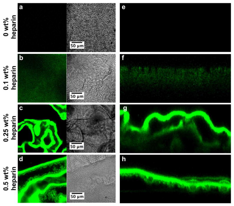
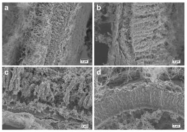
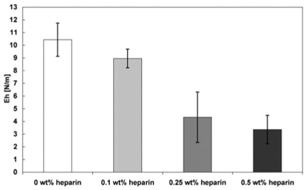
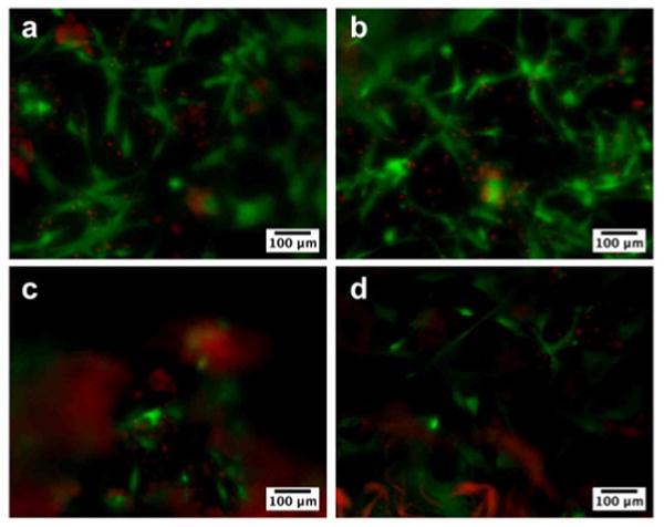
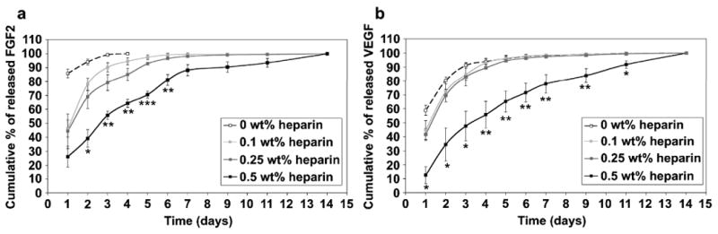
We report here on a bioactive hierarchically structured membrane formed by self-assembly. The membrane is formed with hyaluronic acid and peptide amphiphiles with binding affinity for heparin, and its hierarchical structure contains both an amorphous zone and a layer of fibrils oriented perpendicular to the membrane plane. The design of bioactivity is based on the potential ability to bind and slowly release heparin-binding growth factors. Human mesenchymal stem cells (hMSCs) seeded on these membranes attached and remained viable. Basic fibroblast growth factor (FGF2) and vascular endothelial growth factor (VEGF) were incorporated within the membrane structure prior to self-assembly and released into media over a prolonged period of time (14 days). Using the chicken chorioallantoic membrane (CAM) assay, we also found that these membranes induced a significant and rapid enhancement of angiogenesis relative to controls.


Figures

Figure 1
Figure 1
Confocal laser scanning microscopy images of membranes self-assembled from 2 wt% HBPA and 1 wt% HA with (a, e) 0 wt%, (b, f) 0.1 wt%, (c, g) 0.25 wt%, and (d, h) 0.5 wt% FITC-heparin (green). (a-d) show a representative 1 μm-thick optical slice of each membrane in fluorescence and phase modes. (e-h) are cross-sections of the membrane compiled from the z-stack.
Figure 2
Figure 2
Scanning electron micrographs of membranes self-assembled from 2 wt% HBPA and 1 wt% HA with (a) 0 wt%, (b) 0.1 wt%, (c) 0.25 wt%, and (d) 0.5 wt% heparin incubated for 4 hours. Images show representative cross-sections of the membranes.
Figure 3
Figure 3
Area modulus Eh determined by membrane inflation of membranes self-assembled from 2 wt% HBPA and 1 wt% HA with varying concentrations of heparin after incubating for 4 hours. Increasing heparin concentration correlates to a decrease in area modulus.
Figure 4
Figure 4
Fluorescence microscopy images showing viability of human mesenchymal stem cells (hMSCs) seeded on membranes formed from 2 wt% HBPA and 1 wt% HA with (a) 0 wt%, (b) 0.1 wt%, (c) 0.25 wt%, and (d) 0.5 wt% heparin at day 5. HMSCs were stained for live (green) and dead (red) cells and appeared to attach and grow on all membranes. The difference in cell density is not a reflection of an effect on proliferation but rather a result of uneven resolution because the membranes are not completely flat and become more wrinkled with increasing heparin concentration. Nonspecific red fluorescence was also seen in membranes containing higher amounts of heparin.
Figure 5
Figure 5
Cumulative release of (a) FGF-2 and (b) VEGF from membranes self assembled from 2 wt% HBPA and 1 wt% HA with varying concentrations of heparin. Statistical significance is only shown for 0.5 wt% heparin against all other groups (*P<0.05, **P<0.01, ***P<0.001).
Figure 6
Figure 6
Digital images of membranes self-assembled from 2 wt% HBPA and 1 wt% HA with (a, b) 0 wt% heparin, (c, d) 0 wt% heparin with VEGF and FGF2, (e, f) 0.5 wt% heparin, and (g, h) 0.5 wt% heparin with VEGF and FGF2 on the chorioallantoic membrane on days 0 and 1. Arrows in (h) indicate vessel morphology indicative of VEGF signaling.
Figure 7
Figure 7
Vessel density relative to day 0 induced by HA/HBPA membranes with 0 wt% or 0.5 wt% heparin with or without VEGF and FGF2 (***P<0.001).

References

    1. Webber MJ, Kessler JA, Stupp SI. Emerging peptide nanomedicine to regenerate tissues and organs. J Intern Med. 2010;267(1):71–88. - PMC - PubMed
    1. Langer R, Tirrell DA. Designing materials for biology and medicine. Nature. 2004;428(6982):487–492. - PubMed
    1. Rinaudo M. Main properties and current applications of some polysaccharides as biomaterials. Polym Int. 2008;57(3):397–430.
    1. Baldwin AD, Kiick KL. Polysaccharide-modified synthetic polymeric biomaterials. Peptide Science. 2010;94(1):128–140. - PMC - PubMed
    1. Silva GA, Czeisler C, Niece KL, Beniash E, Harrington DA, Kessler JA, Stupp SI. Selective differentiation of neural progenitor cells by high-epitope density nanofibers. Science. 2004;303(5662):1352–1355. - PubMed

Publication types

MeSH terms