Review
. 2011 Feb;54(2):366-73.
doi: 10.1016/j.jhep.2010.10.003. Epub 2010 Oct 14.

Hedgehog signaling in the liver

Affiliations
Review

Hedgehog signaling in the liver

Alessia Omenetti et al. J Hepatol. 2011 Feb.

Abstract

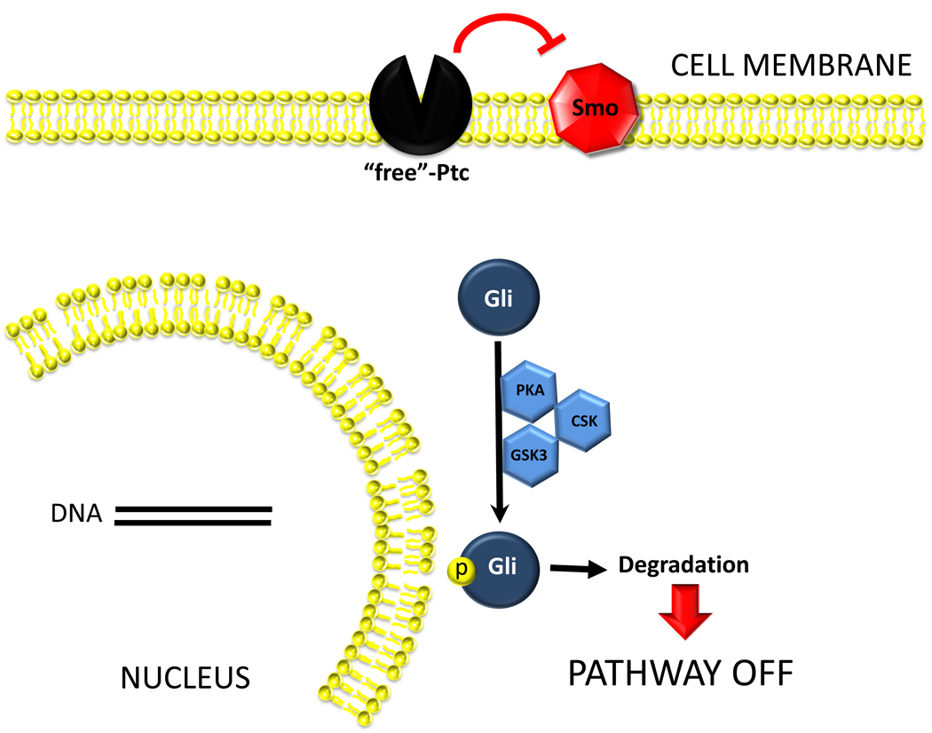
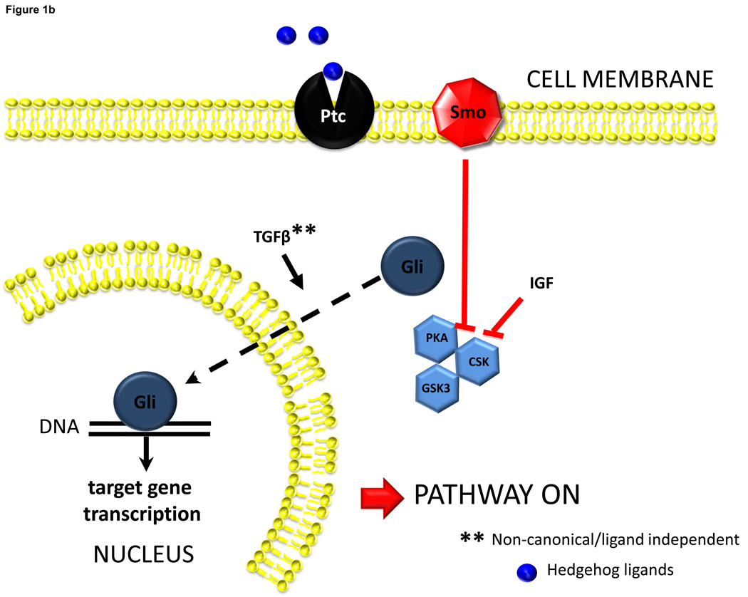
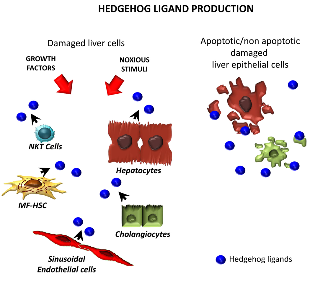
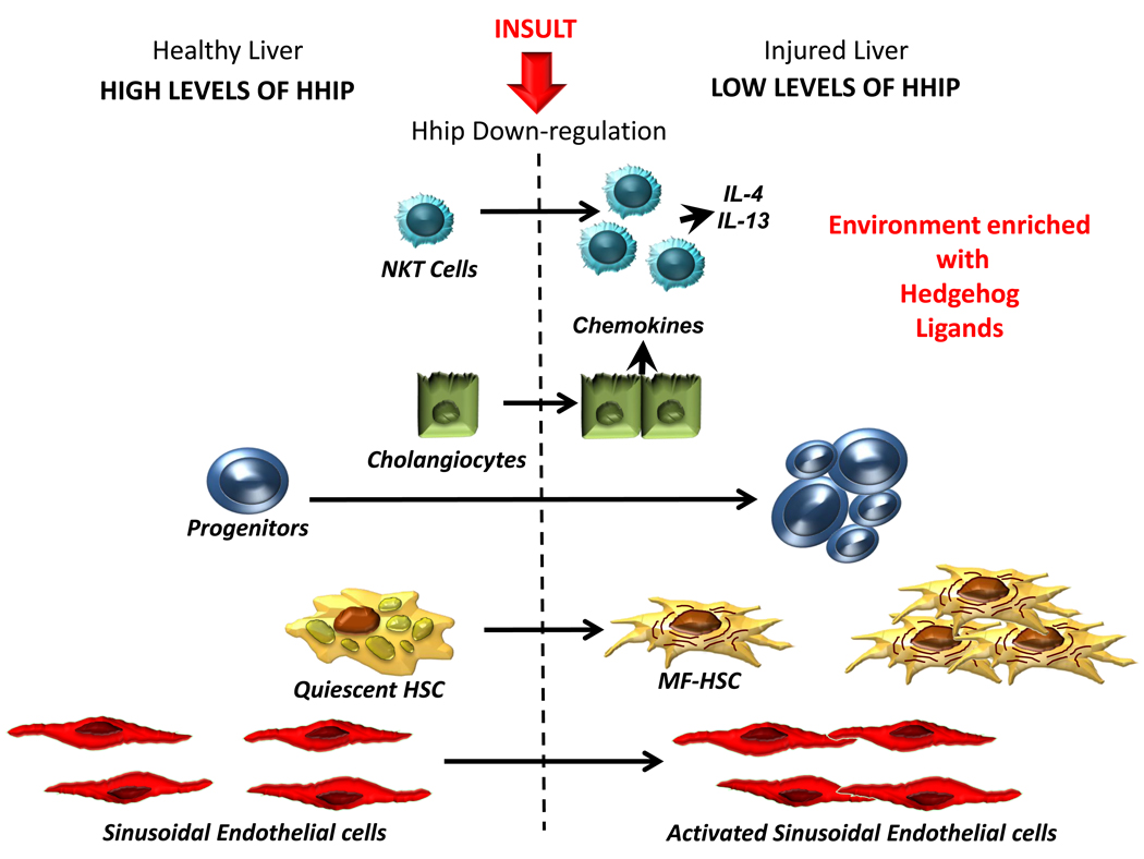
Reactivation of Hedgehog (Hh), a morphogenic signaling pathway that controls progenitor cell fate and tissue construction during embryogenesis occurs during many types of liver injury in adult. The net effects of activating the Hedgehog pathway include expansion of liver progenitor populations to promote liver regeneration, but also hepatic accumulation of inflammatory cells, liver fibrogenesis, and vascular remodeling. All of these latter responses are known to be involved in the pathogenesis of cirrhosis. In addition, Hh signaling may play a role in primary liver cancers, such as cholangiocarcinoma and hepatocellular carcinoma. Study of Hedgehog signaling in liver cells is in its infancy. Additional research in this area is justified given growing experimental and clinical data supporting a role for the pathway in regulating outcomes of liver injury.


Figures

Figure 1
Figure 1
Figure 1a. Hh pathway is silent in Hh-responsive cells when Hh ligands are absent. Cells that are capable of responding to Hh ligands (i.e., Hh-responsive cells) express Hh receptors. Patched (Ptc) is the receptor that physically interacts with Hh ligands. In the absence of Hh ligands, Ptc represses the activation of a co-receptor-like molecule, Smoothened (Smo). This repression prevents Smo from interacting with other intracellular factors that permit the stabilization and accumulation of Glioblastoma (Gli) transcription factors. Thus, Gli proteins undergo phosphorylation by various intracellular kinases (PKA, GSK3b, CSK), become ubiquitinated, move to proteasomes and are degraded. Reduced availability of Gli factors influences the transcription of their target genes. Lack of Gli1 and Gli2 generally reduces target gene transcription, while lack of Gli3 can either stimulate or inhibit transcriptional activity. Figure 1b. Hh ligands activate Hh pathway signaling. Interaction between Hh ligands and Ptc liberates Smoothened from the normal repressive actions of Ptc. This results in eventual inhibition of factors the promote Gli phosphorylation/degradation, and permits cellular accumulation of Gli. Other factors that inhibit Gli-phosphorylation, such as insulin like growth factor-1 (IGF), have also been shown to facilitate stabilization of Gli1 in cells that are otherwise capable of producing this protein. There is also a report that Transforming Growth Factor beta (TGFb) can stimulate Gli accumulation via mechanisms that may operate independently of Smoothened. Nuclear accumulation of Gli factors, in turn, influences transcriptional activity of Gli-target genes. Gli1 and Gli2 generally increase gene transcription, while Gli3 can either increase or decrease gene transcription depending on its post-translational modification.
Figure 1
Figure 1
Figure 1a. Hh pathway is silent in Hh-responsive cells when Hh ligands are absent. Cells that are capable of responding to Hh ligands (i.e., Hh-responsive cells) express Hh receptors. Patched (Ptc) is the receptor that physically interacts with Hh ligands. In the absence of Hh ligands, Ptc represses the activation of a co-receptor-like molecule, Smoothened (Smo). This repression prevents Smo from interacting with other intracellular factors that permit the stabilization and accumulation of Glioblastoma (Gli) transcription factors. Thus, Gli proteins undergo phosphorylation by various intracellular kinases (PKA, GSK3b, CSK), become ubiquitinated, move to proteasomes and are degraded. Reduced availability of Gli factors influences the transcription of their target genes. Lack of Gli1 and Gli2 generally reduces target gene transcription, while lack of Gli3 can either stimulate or inhibit transcriptional activity. Figure 1b. Hh ligands activate Hh pathway signaling. Interaction between Hh ligands and Ptc liberates Smoothened from the normal repressive actions of Ptc. This results in eventual inhibition of factors the promote Gli phosphorylation/degradation, and permits cellular accumulation of Gli. Other factors that inhibit Gli-phosphorylation, such as insulin like growth factor-1 (IGF), have also been shown to facilitate stabilization of Gli1 in cells that are otherwise capable of producing this protein. There is also a report that Transforming Growth Factor beta (TGFb) can stimulate Gli accumulation via mechanisms that may operate independently of Smoothened. Nuclear accumulation of Gli factors, in turn, influences transcriptional activity of Gli-target genes. Gli1 and Gli2 generally increase gene transcription, while Gli3 can either increase or decrease gene transcription depending on its post-translational modification.
Figure 2
Figure 2
Figure 2a. Differential Activity of Hedgehog Pathway in Healthy and Injured Livers. Healthy livers express low levels of Hedgehog (Hh) ligands. Several types of resident liver cells are capable of producing Hh ligands, including hepatocytes, cholangiocytes, hepatic stellate cells (HSC), natural killer T (NKT) cells, and sinusoidal endothelial cells. Ligand production can be stimulated by growth factors/cytokines, as well as by cytotoxic/apoptotic stress. Thus, diverse stimuli that promote liver regeneration/remodeling induce hepatic production of Hh ligands. Figure 2b. Differential Activity of Hedgehog Pathway in Healthy and Injured Livers. Healthy livers express low levels of Hh ligands (a) and relatively high levels of Hh interacting protein (Hhip) (b), which binds to Hh ligands, preventing them from engaging receptors on Hh-responsive target cells. During liver injury, production of Hh ligands increases (Fig 2a) and Hhip is repressed, permitting ligand-receptor interaction and activation of the Hh signaling pathway in Hh-responsive cells. The latter include several types of resident liver cells, including NKT cells, cholangiocytes, progenitors and quiescent hepatic stellate cells (Q-HSC). Activation of Hh signaling in each of these cell types induces responses that contribute to fibrogenic repair. For example, Hh pathway activation stimulates growth of NKT cell populations and induces their production of pro-fibrogenic factors, such as IL4 and IL13. It also stimulates cholangiocyte growth and production of chemokines, including chemokines that recruit NKT cells and other inflammatory/immune cells to the liver. In addition, Hh ligands promote growth of liver progenitors and stimulate Q-HSC to transition to become myofibroblastic (MF)-HSC. The growth of MF-HSC is further stimulated by Hh pathway activity. Coupled with expansion of Hh-responsive ductular and progenitor populations, this contributes to the fibroductular reaction that often accompanies liver injury. Finally, Hh ligands activate liver sinusoidal endothelial cells, causing them to express adhesion factors and other mediators that contribute to vascular remodeling.
Figure 2
Figure 2
Figure 2a. Differential Activity of Hedgehog Pathway in Healthy and Injured Livers. Healthy livers express low levels of Hedgehog (Hh) ligands. Several types of resident liver cells are capable of producing Hh ligands, including hepatocytes, cholangiocytes, hepatic stellate cells (HSC), natural killer T (NKT) cells, and sinusoidal endothelial cells. Ligand production can be stimulated by growth factors/cytokines, as well as by cytotoxic/apoptotic stress. Thus, diverse stimuli that promote liver regeneration/remodeling induce hepatic production of Hh ligands. Figure 2b. Differential Activity of Hedgehog Pathway in Healthy and Injured Livers. Healthy livers express low levels of Hh ligands (a) and relatively high levels of Hh interacting protein (Hhip) (b), which binds to Hh ligands, preventing them from engaging receptors on Hh-responsive target cells. During liver injury, production of Hh ligands increases (Fig 2a) and Hhip is repressed, permitting ligand-receptor interaction and activation of the Hh signaling pathway in Hh-responsive cells. The latter include several types of resident liver cells, including NKT cells, cholangiocytes, progenitors and quiescent hepatic stellate cells (Q-HSC). Activation of Hh signaling in each of these cell types induces responses that contribute to fibrogenic repair. For example, Hh pathway activation stimulates growth of NKT cell populations and induces their production of pro-fibrogenic factors, such as IL4 and IL13. It also stimulates cholangiocyte growth and production of chemokines, including chemokines that recruit NKT cells and other inflammatory/immune cells to the liver. In addition, Hh ligands promote growth of liver progenitors and stimulate Q-HSC to transition to become myofibroblastic (MF)-HSC. The growth of MF-HSC is further stimulated by Hh pathway activity. Coupled with expansion of Hh-responsive ductular and progenitor populations, this contributes to the fibroductular reaction that often accompanies liver injury. Finally, Hh ligands activate liver sinusoidal endothelial cells, causing them to express adhesion factors and other mediators that contribute to vascular remodeling.

Similar articles

Cited by

References

    1. Acloque H, Adams MS, Fishwick K, Bronner-Fraser M, Nieto MA. Epithelial-mesenchymal transitions: the importance of changing cell state in development and disease. J Clin Invest. 2009;119(6):1438–1449. - PMC - PubMed
    1. Adolphe C, Narang M, Ellis T, Wicking C, Kaur P, Wainwright B. An in vivo comparative study of sonic, desert and Indian hedgehog reveals that hedgehog pathway activity regulates epidermal stem cell homeostasis. Development. 2004;131(20):5009–5019. - PubMed
    1. Alinger B, Kiesslich T, Datz C, Aberger F, Strasser F, Berr F, et al. Hedgehog signaling is involved in differentiation of normal colonic tissue rather than in tumor proliferation. Virchows Arch. 2009;454(4):369–379. - PubMed
    1. Alvaro D, Mancino MG. New insights on the molecular and cell biology of human cholangiopathies. Mol Aspects Med. 2008;29(1–2):50–57. - PubMed
    1. Baum B, Settleman J, Quinlan MP. Transitions between epithelial and mesenchymal states in development and disease. Semin Cell Dev Biol. 2008;19(3):294–308. - PubMed

Publication types

Substances