. 2010 Nov 24;33(5):817-29.
doi: 10.1016/j.immuni.2010.10.018.

Endothelial heparan sulfate controls chemokine presentation in recruitment of lymphocytes and dendritic cells to lymph nodes

Affiliations

Endothelial heparan sulfate controls chemokine presentation in recruitment of lymphocytes and dendritic cells to lymph nodes

Xingfeng Bao et al. Immunity. .

Erratum in

  • Immunity. 2011 May 27;34(5):820. Petryanik, Bronislawa [corrected to Petryniak, Bronislawa]

Abstract

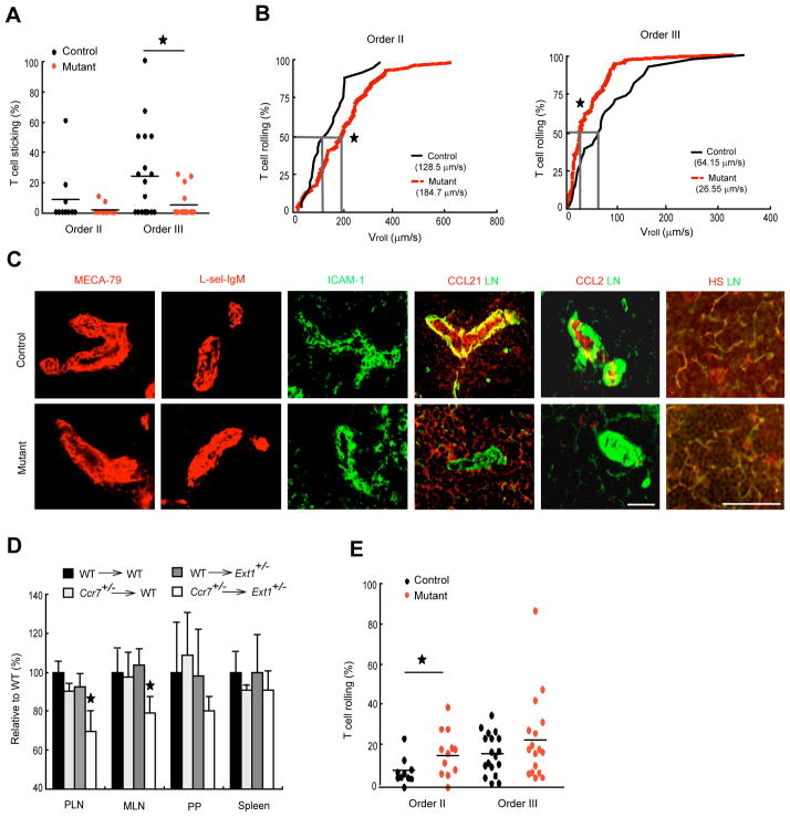
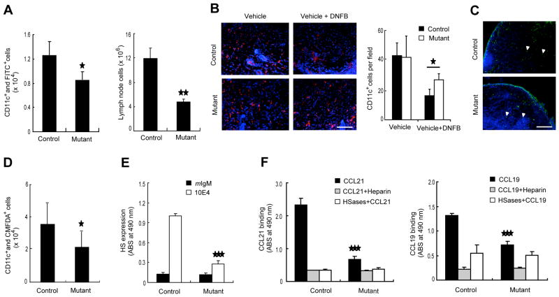
Heparan sulfate can bind several adhesion molecules involved in lymphocyte trafficking. However, the in vivo function of endothelial heparan sulfate in lymphocyte homing and stimulation of the immune response has not been elucidated. Here, we generated mutant mice deficient in the enzyme Ext1, which is required for heparan sulfate synthesis, in a Tek-dependent and inducible manner. Chemokine presentation was diminished in the mutant mice, causing the lack of appropriate integrin-mediated adhesion, and resulted in a marked decrease in lymphocyte sticking to high endothelial venules and in recruitment of resident dendritic cells through lymphatic vessels to the lymph nodes. As a consequence, mutant mice displayed a severe impairment in lymphocyte homing and a compromised contact hypersensitivity response. By contrast, lymphocyte rolling was increased because of loss of electrostatic repulsion by heparan sulfate. These results demonstrate critical roles of endothelial heparan sulfate in immune surveillance and immune response generation.


Figures

Figure 1
Figure 1
Diminished lymphocyte homing upon enzymatic removal of endothelial heparan sulfate. (A) Homing of labeled WT lymphocytes to lymph nodes of enzyme-infused WT mice. Chondroitinase ABC (CSase ABC) or a mixture of heparitinase and heparinase (HSases) in PBS was injected through mouse tail veins 2 hr before cell injection (n = 3–5). PBS was infused as a control. *, P < 0.05; ***, P < 0.001, versus PBS. (B) Immunostaining of lymph node frozen sections. Monoclonal antibodies (mAbs) 10E4 and 3G10 detected intact heparan sulfate and digested HS proteoglycans (ΔHS), respectively, while mAbs CS56 and 2B6 stained chondroitin sulfate and digested CS proteoglycans (ΔCS), respectively. Bar, 50 μm. (C) Homing of HSase-treated and labeled lymphocytes from WT mice to the lymph nodes of WT mice. PBS treatment was run as a control. (D) RT-PCR analysis of membrane-associated heparan sulfate proteoglycans in isolated MECA-79+ cells from WT mice and a control mouse myoblast cell line C2C12. (E) Immune blot of lymph node vascular heparan sulfate proteoglycans. Matrix of lymph nodes from the WT mice that received Hsases and CSase ABC was solubilized with Triton X-100-containing buffer, separated by SDS-PAGE and blotted with mAb 3G10. Equal amounts of proteins were loaded on each lane. The migration position of perlecan, glypican-1 (Gpn1) and syndecan-4 (Syn4) are indicated. Data in A and C are representative of four independent experiments (mean ± S.E.).
Figure 2
Figure 2
Inducible abrogation of endothelial heparan sulfate in Ext1 mutant mice. (A) Immunostaining of high endothelial venule (HEV) heparan sulfate in frozen lymph node (LN) sections by mAb 10E4. Co-staining with CD31 or perlecan was performed. Bar, 50 μm. (B) Analysis of PCR products of Ext1 in HEV cells isolated from the control and mice. ΔExt1 denotes deficient Ext1 gene. (C) Immune blot of syndecan-4 (Syn-4) in lung endothelial cells isolated from control and mutant mice. Pretreatment with a mixture of heparitinase and heparinase (Hsases) and/or chondroitinase ABC (CSase ABC) at 37°C for 1 hr was included. (D) ELISA of heparan sulfate chains on syndencan-4 or control (IgG) immunoprecipitate from lung endothelial cells of the control and mutant mice by using mAb 10E4. Data are expressed as mean ± S.D..
Figure 3
Figure 3
Impaired lymphocyte homing and lymph node cellularity in Ext1 mutant mice. (A) Cell numbers of secondary lymphoid organs in mice that received doxcycline for 3–4 weeks (n = 5–9). (B) Numbers of white blood cells (WBC) and subtypes in the peripheral blood of the mice after 4 weeks treatment with doxycyline (n = 8). (C) Cell numbers of PLNs in the mice that received doxcycline for 2 months (n = 4–6). Insert shows the visual size of the representative inguinal lymph nodes from both mouse lines. Bar, 0.5 cm. (D) Proliferation of lymph node cells stimulated by Concanavalin A at a dose of 2 μg/ml. (E) Populations of T cells (CD3+) and B cells (B220+) in secondary lymphoid organs. (F) Homing of labeled lymphocytes from WT mice to the secondary lymphoid organs of the control and the mutant mice (n = 3–5). An infusion of heparitinase and heparinase (HSases) was included for the mutant mice. (G) Homing of labeled lymphocytes isolated from the control or the mutant mice to the lymph nodes of WT mice (n = 3–5). (H) Immunostaining of lymph node sections with anti-CD3 (in red) and anti-B220 (in green). Bar, 200 μm. *, P < 0.05; **, P < 0.01, versus controls. Data are representative of two or three independent experiments (mean ± S.E.).
Figure 4
Figure 4
Diminished sticking and decreased rolling velocity of lymphocytes in Ext1 mutant mice. (A) Sticking fraction of T cells in orders II and III lymph node venules analyzed by intravital microscopy. Combined results of 6–7 venules for order II and 13 venules for order III from 8 mice of each group are shown. (B) Cumulative rolling velocity of T cells in orders II and III lymph node venules of control and mutant mice (n = 8). The percentage of rolling cells is expressed as function of Vroll (see Experimental Procedures). (C) Immunostaining of MECA-79 antigen (MECA-79), L-selectin ligands (L-sel-IgM), intercellular adhesion molecule-1 (ICAM-1), chemokines CCL21 and CCL2, and laminin (LN) in lymph node frozen sections. Bars, 50 μm. (D) Homing of labeled lymphocytes isolated from WT or Ccr7+/− mice to the lymph nodes of WT mice or Ext1+/ mice (n = 4). (E) Rolling fraction of T cells in orders II and III lymph node venules analyzed by intravital microscopy. Combined results of 6–7 venules for order II and 13 venules for order III from 8 mice of each group are shown. Mean values are shown as horizontal bars in A and E. *, P < 0.05, versus control mice (unpaired t test).
Figure 5
Figure 5
Rolling of 38C13 cells on CHO cells expressing 6-sulfo sLeX or sLeX on N-glycans and O-glycans. (A) Rolling of 38C13 cells on CHO cells expressing 6-sulfo sLeX on both N- and O-glycans (CHO:CD34:FucT-VII:C1:C2:GlcNAc6ST-2) or on CHO cells expressing only N-glycans sLeX (CHO:CD34:FucT-VII) without enzyme treatment (−) or treated with chondroitinase ABC (CSase ABC), heparitinase and heparinase (HSases) (+). (B) Cell rolling on CHO cells expressing 6-sulfo sLeX on both N- and O-glycans treated with individual enzyme described in A. (C) Rolling velocity on PBS- or HSase-treated CHO cells expressing 6-sulfo sLeX as shown in B. Data are representative of two independent experiments (mean ± S.E.).
Figure 6
Figure 6
Impaired contact hypersensitivity in Ext1 mutant mice. (A) Ear swelling 24 hr after challenge with DNFB or vehicle alone in control and mutant mice (n = 4–6). (B) Hematoxylin and eosin staining of ear sections challenged with DNFB. Arrowheads are infiltrated mononuclear cells; arrows are infiltrated neutrophils; dashed areas are epidermal areas with neutrophil clustering. Bar, 250 μm. (C) Quantitation of infiltrated T cells in inflamed ears (left panels) and areas of neutrophil clusters (right panels) (n = 3–5). Numbers of CD3ε+ cells in the area of 300 μm long ear were counted, while the epidermis area with clustered neutrophils of the whole ear were measured. (D) Cell number of sensitized draining inguinal lymph nodes (left panels) and lymphocyte homing to the draining inguinal lymph nodes (right panels) (n= 3–5). (E) CXCL2 expression in CD31+ cells of DNFB-inflamed abdominal skins. Arrowheads indicate microvessels. Bar, 25 μm. (F) Cell proliferation isolated from sensitized inguinal lymph nodes in response to DNBS (200 μg/ml) (n= 3). *, P < 0.05; **, P < 0.01, versus control mice. Data are representative of two independent experiments (mean ± S.E.)
Figure 7
Figure 7
Impaired migration of dendritic cells to lymph nodes in Ext1 mutant mice. (A) Cell numbers of draining lymph nodes (right panels) and homing of dendritic cells to draining lymph nodes (left panels) in skin painting experiment (n = 4–5). (B) Immunofluorescent staining of resident dendritic cells (red) in DNFB-inflamed skin epidermis. Nuclei (blue) were stained by Hoechst. Bar, 100 μm. (C) Immunofluorescence of draining lymph nodes in skin painting experiments. Arrowheads point to infiltrated FITC-bearing dendritic cells. Bar, 200 μm. (D) Homing of intrademally loaded dendritic cells to draining lymph nodes (n = 3–5). (E) ELISA of heparan sulfate expression assessed by mAb 10E4 in cultured mouse skin lymphatic endothelial cells. (F) Binding of recombinant mouse CCL21 or CCL19 to cultured skin lymphatic endothelial cells. In some cases, addition of heparin (100 μg/ml) or a pretreatement with heparitinase and heparinase (HSases) at 37 °C for 1 h was included. *, P < 0.05; **, P < 0.01; ***, P < 0.001 versus control mice. Data are expressed as mean ± S.E. for A, B and D, and mean ± S.D. for E and F.

Comment in

Similar articles

Cited by

References

    1. Arbones ML, Ord DC, Ley K, Ratech H, Maynard-Curry C, Otten G, Capon DJ, Tedder TF. Lymphocyte homing and leukocyte rolling and migration are impaired in L-selectin-deficient mice. Immunity. 1994;1:247–260. - PubMed
    1. Bajenoff M, Egen JG, Koo LY, Laugier JP, Brau F, Glaichenhaus N, Germain RN. Stromal cell networks regulate lymphocyte entry, migration, and territoriality in lymph nodes. Immunity. 2006;25:989–1001. - PMC - PubMed
    1. Belteki G, Haigh J, Kabacs N, Haigh K, Sison K, Costantini F, Whitsett J, Quaggin SE, Nagy A. Conditional and inducible transgene expression in mice through the combinatorial use of Cre-mediated recombination and tetracycline induction. Nucleic Acids Res. 2005;33:e51. - PMC - PubMed
    1. Bishop JR, Schuksz M, Esko JD. Heparan sulphate proteoglycans fine-tune mammalian physiology. Nature. 2007;446:1030–1037. - PubMed
    1. Butcher EC, Picker LJ. Lymphocyte homing and homeostasis. Science. 1996;272:60–66. - PubMed

Publication types

MeSH terms