. 2010 Nov 24;33(5):713-22.
doi: 10.1016/j.immuni.2010.11.010.

MicroRNAs prevent the generation of autoreactive antibodies

Affiliations

MicroRNAs prevent the generation of autoreactive antibodies

Laura Belver et al. Immunity. .

Abstract

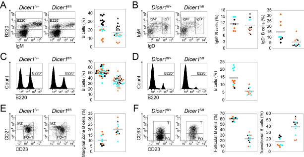
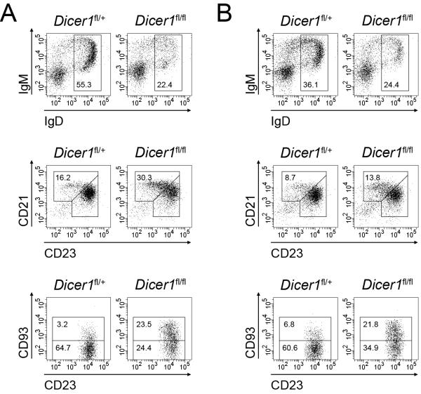
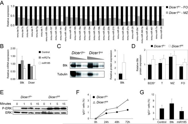
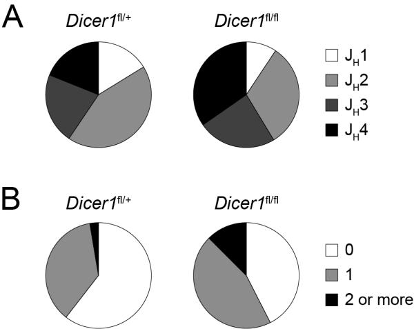
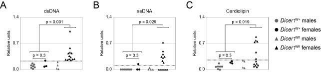
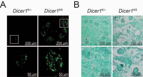
MicroRNAs have been shown to be critical for a number of aspects of immune system regulation and function. Here, we have examined the role of microRNAs in terminal B cell differentiation by analyzing Cd19-Cre(ki/+) Dicer1(fl/fl) mice. We found that in the absence of Dicer, the transitional and marginal zone (MZ) B cell compartments were overrepresented and follicular (FO) B cell generation was impaired. microRNA analysis revealed that miR185, a microRNA overexpressed in FO cells, dampened B cell receptor (BCR) signaling through Bruton tyrosine kinase downregulation. Dicer-deficient B cells had a skewed BCR repertoire with hallmarks of autoreactivity, which correlated with high titers of autoreactive antibodies in serum and autoimmune features in females. Together, our results reveal a crucial role for microRNAs in late B cell differentiation and in the establishment of B cell tolerance.


Figures

Figure 1
Figure 1. Total peripheral B cells are reduced and MZ and T subsets are overrepresented in the absence of Dicer
Phenotypic analysis of bone marrow (A and B), spleen (C, E and F) and lymph nodes (D) from CD19-Creki/+Dicerfl/+ and CD19-Creki/+Dicerfl/fl mice. Cell suspensions were stained with the indicated antibodies and analysed by flow cytometry. Representative FACS analyses are shown on the left for total cells (A, C and D), B220+-gated cells (B and E) or B220+CD23bright-gated cells (F). Graphs on the right show the frequency of the indicated subsets in individual mice analysed as follows, (A) B220+ bone marrow B cells (n=18-21, p=0.01); (B) IgM+IgD immature B (n=8-11, p=0.62) and IgD+ recirculating B cells (n=8-11, p<0.01); (C) B220+ spleen B cells (n=31-33, p<0.01); (D) B220+ lymph node cells (n=11, p<0.01); (E) CD21brightCD23+ marginal zone (MZ) (n=9-11, p<0.01) and (F) CD21+CD23brightCD93 follicular (FO) (n=9-11, p<0.01) and CD21+CD23brightCD93+ transitional (T) cells (n=9-11, p<0.01) within total spleen B cells. Circles represent CD19-Creki/+Dicerfl/+ mice and triangles represent CD19-Creki/+Dicerfl/fl mice. Color code represents mouse age (black, <10 weeks old; blue, 10 to 20 weeks old and orange, >20 weeks old). Mean values are shown as horizontal bars. See also Figure S1 and Table S1.
Figure 2
Figure 2. Dicer deficient cells in mixed chimeras show a reduction in total peripheral B cell generation and an overrepresentation of MZ and T subsets
Phenotypic analysis of bone marrow and spleen from lethally irradiated mice 12 weeks after bone marrow transfer with 100% CD45.2+ CD19-Creki/+Dicerfl/+ or 100% CD45.2+ CD19-Creki/+Dicerfl/fl cells (A) and 1:1 mixtures of bone marrow cells from CD45.2+ CD19-Creki/+Dicerfl/+ or CD45.2+ CD19-Creki/+Dicerfl/fl mice with bone marrow cells from CD45.1+ wild type mice (B). Representative FACS analyses for the indicated markers are shown for B220+-gated CD45.2+ bone marrow cells (top histograms), B220+-gated CD45.2+ spleen cells (middle histograms) and B220+CD23bright-gated CD45.2+ spleen cells (bottom histograms). Numbers in the gates show the percentage mean within CD45.2+B220+ cells of the following populations: top histograms, IgD+ recirculating B cells (A: n=3, p<0.01; B: n=4, p<0.01); middle histograms, CD21brightCD23+ marginal zone B cells (A: n=3, p<0.01; B: n=4, p=0.03); bottom histograms, CD21+CD23brightCD93 follicular B cells (A: n=3, p<0.01; B: n=4, p<0.01) and CD21+CD23brightCD93+ transitional B cells (A: n=3, p<0.01; B: n=4, p<0.01). Total numbers, means and standard deviations of the indicated cell subsets in the mixed chimeras are shown in Table S2. See also Figure S2.
Figure 3
Figure 3. microRNA analysis of FO and MZ B cells and BCR signalling alterations in Dicer deficient cells
(A) microRNA profiling of FO and MZ subsets of CD19-Creki/+Dicerfl/+ mice. CD21+CD23bright follicular (FO) and CD21brightCD23+ marginal zone (MZ) B cells were separated by cell sorting from CD19-Creki/+Dicerfl/+ spleens. RNA was isolated, labelled, and hybridized on Agilent microRNA arrays. Differentially expressed microRNAs in MZ (grey bars) vs FO (black bars) cells (see Methods) are shown (p<0.1). Bars represent fluorescence intensity normalized to FO cells (mean values of 3 independent experiments). (B) Differentially expressed microRNAs (shown in A) were subjected to target prediction analysis of genes potentially involved in MZ vs FO cell differentiation and/or maintenance. miR185 was predicted to target Btk by all three miRNA target prediction softwares used (MiRanda, TargetScan and miRBase). Control, miR185 and miR27a (not predicted to target Btk) retroviral vectors were transduced into primary B cells from wild type spleens in the presence of LPS and IL4. Two days after transduction RNA was isolated and Btk levels were measured by real-time RT-PCR (control vs miR185: n=4, p<0.01). Dicer mRNA levels from the same samples are shown as a negative control (control vs miR185: n=4, p=0.73). Bars represent mRNA levels after normalization to GAPDH expression and relative to control-transduced cells. Standard deviations are shown. (C) Btk protein level is increased in spleen B cells of Dicer deficient mice. 2-fold serial dilutions of total lysates from CD19-Creki/+Dicerfl/+ and CD19-Creki/+Dicerfl/fl spleen B cells were analysed by Western blotting with anti-Btk and anti-tubulin antibodies. Densitometric quantification is shown on the right, black bar represents CD19-Creki/+Dicerfl/+ mice and white bar represents CD19-Creki/+Dicerfl/fl mice (n=2, p<0.01). (D) Btk protein expression in B cell subsets. Spleen cell suspensions from CD19-Creki/+Dicerfl/+ (black bars) and CD19-Creki/+Dicerfl/fl mice (white bars) were stained with anti-B220, anti-CD21, anti-CD23 and anti-CD93, intracellular stained with anti-Btk antibody and analysed by flow cytometry. Mean fluorescence in the indicated subsets was normalized to CD19-Creki/+Dicerfl/+ cell fluorescence in total B220+ cells (B220+: n=8-10, p<0.01; T: n=8-10, p=0.02; MZ: n=8-10, p=0.41; FO: n=8-10, p<0.01). (E) Increased phospho-ERK levels in CD19-Creki/+Dicerfl/fl cells. CD19-Creki/+Dicerfl/+ and CD19-Creki/+Dicerfl/fl spleen B cells were stimulated with anti-IgM and total lysate from 1x10⁶ cells was loaded per lane for analyzing by Western blotting anti-phospho-ERK and anti-ERK protein levels. Time after stimulation is indicated over each lane. (F) Increased class switching in Dicer deficient cells upon stimulation with anti-IgM. Spleen B cells from CD19-Creki/+Dicerfl/+ (filled circles) and CD19-Creki/+Dicerfl/fl mice (triangles) were isolated and cultured in the presence of anti-IgM and IL4. The efficiency of class switch recombination to IgG1 was analysed by flow cytometry at the indicated time points (n=3). (G) Btk and miR185 gain-of-function analysis. Wild type spleen B cells were transduced with Btk, miR185 or empty vectors in the presence of anti-IgM and IL4. The efficiency of class switching to IgG1 was analysed by flow cytometry 96 hours after transduction. Bars represent the percentage of IgG1+ cells within transduced GFP+ cells. Standard deviations from three independent experiments are shown (n=3, p=0.03). See also Figure S3.
Figure 4
Figure 4. CD19-Creki/+Dicerfl/fl B cells express a skewed Ig repertoire
Analysis of the expressed IgH repertoire in control and Dicer deficient B cells. B cells were isolated from CD19-Creki/+Dicerfl/+ and CD19-Creki/+Dicerfl/fl spleens by immunomagnetic depletion. RNA was extracted, retrotranscribed and PCR-amplified with oligonuclotides specific for VHJ558 or VH7183 in combination with a Cμ oligonucleotide. PCR products were cloned, sequenced and analysed using IgBLAST software. Sectors represent the contribution of JH segments (A) (n=74-75, p<0.01) and of sequences containing 0, 1 or >=2 R+K residues at CDR3 (B) (n=74-75, p=0.03) for CD19-Creki/+Dicerfl/+ and CD19-Creki/+Dicerfl/fl B cells. Results were pooled from 4 independent animals of each genotype. Complete sequence analysis is shown in Table S3.
Figure 5
Figure 5. CD19-Creki/+Dicerfl/fl mice have high serum titers of autoreactive antibodies
Analysis of serum Ig titers against self-antigens. Serum from 40-60 week old CD19-Creki/+Dicerfl/+ (circles) and CD19-Creki/+Dicerfl/fl (triangles) animals was collected and reactivity against dsDNA (A), ssDNA (B) and cardiolipin (C) was assessed by ELISA. The results are represented as relative colorimetric units. Background signal from RAG knock-out mouse serum was substracted and values were normalized to the signal obtained from MRLlpr/lpr mouse serum. For clarity, results obtained from males (grey) and females (black) are represented separately. A threshold for autoreactive antibody titers was established by adding two standard deviations to the mean value of the titers detected in the control CD19-Creki/+Dicerfl/+ mice (shown as a grey dotted line). P values (Fisher’s exact test) between the indicated groups are shown (n=9-17). See also Figure S4.
Figure 6
Figure 6. CD19-Creki/+Dicerfl/fl females display autoimmune features
(A) Immunocomplexes in kidneys from Dicer deficient females. Kidney sections from 40-60 week old CD19-Creki/+Dicerfl/+ and CD19-Creki/+Dicerfl/fl animals were stained with anti-IgG antibodies. Representative immunofluorescence glomerular stainings are shown at two different magnifications. Scale bars are indicated. (B) Kidney damage in CD19Creki/+Dicerfl/fl mice. Formalin-fixed kidney sections from CD19Creki/+Dicerfl/+ and CD19Creki/+Dicerfl/fl aged mice were subjected to Silver-PAS staining. Scale bars are shown. See also Figure S5.

Similar articles

Cited by

References

    1. Allman D, Pillai S. Peripheral B cell subsets. Curr Opin Immunol. 2008;20:149–157. - PMC - PubMed
    1. Bassing CH, Swat W, Alt FW. The mechanism and regulation of chromosomal V(D)J recombination. Cell. 2002;109(Suppl):S45–55. - PubMed
    1. Carsetti R, Rosado MM, Wardmann H. Peripheral development of B cells in mouse and man. Immunol Rev. 2004;197:179–191. - PubMed
    1. Crouzier R, Martin T, Pasquali JL. Heavy chain variable region, light chain variable region, and heavy chain CDR3 influences on the mono- and polyreactivity and on the affinity of human monoclonal rheumatoid factors. J Immunol. 1995;154:4526–4535. - PubMed
    1. Dammers PM, Visser A, Popa ER, Nieuwenhuis P, Kroese FG. Most marginal zone B cells in rat express germline encoded Ig VH genes and are ligand selected. J Immunol. 2000;165:6156–6169. - PubMed

Publication types

Associated data