Review
. 2011 Feb;93(2):244-69.
doi: 10.1016/j.pneurobio.2010.11.001. Epub 2010 Nov 18.

Neuronal control of swimming behavior: comparison of vertebrate and invertebrate model systems

Affiliations
Review

Neuronal control of swimming behavior: comparison of vertebrate and invertebrate model systems

Olivia J Mullins et al. Prog Neurobiol. 2011 Feb.

Abstract

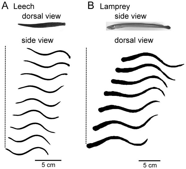
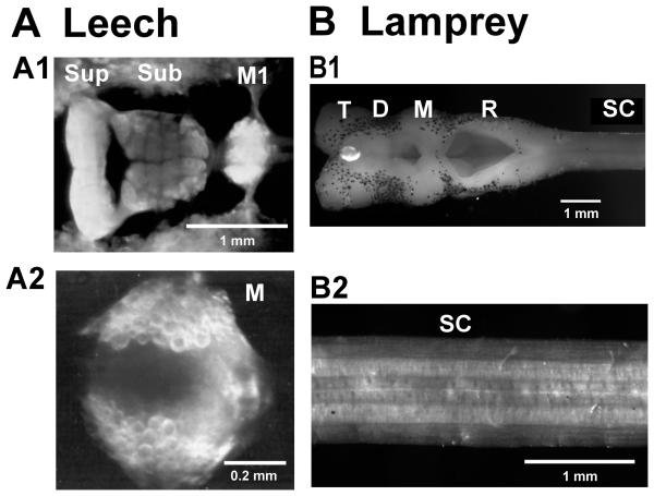
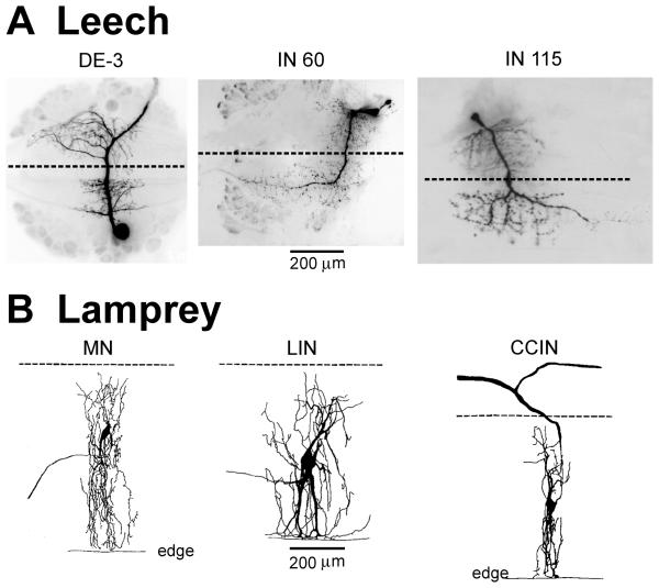
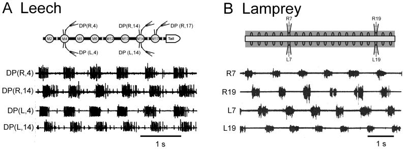
Swimming movements in the leech and lamprey are highly analogous, and lack homology. Thus, similarities in mechanisms must arise from convergent evolution rather than from common ancestry. Despite over 40 years of parallel investigations into this annelid and primitive vertebrate, a close comparison of the approaches and results of this research is lacking. The present review evaluates the neural mechanisms underlying swimming in these two animals and describes the many similarities that provide intriguing examples of convergent evolution. Specifically, we discuss swim initiation, maintenance and termination, isolated nervous system preparations, neural-circuitry, central oscillators, intersegmental coupling, phase lags, cycle periods and sensory feedback. Comparative studies between species highlight mechanisms that optimize behavior and allow us a broader understanding of nervous system function.


Figures

Figure 1
Figure 1
Block diagram of leech and lamprey systems that control swimming. Arrows indicate the bidirectionality of all interactions but swim initiation.
Figure 2
Figure 2
Body undulations in swimming leeches and lampreys. A Video frames of a swimming leech (Hirudo verbana). Dorsal view show shows the elongated body from above; side view shows the body undulations. Profiles were captured at 100 fps, with every fourth frame shown, for one complete cycle. The dashed reference line indicates forward progression during the 0.35 s cycle period. B Video frames of a lamprey (Petromyzon marinus; young adult). Side view shows the body profile from the side, at rest; dorsal view shows the swimming undulations viewed from above. Swimming profiles were captured at 30 fps, with every second frame shown. The dashed reference line indicates forward progression during the 0.4 s cycle period. Rostral is to the left.
Figure 3
Figure 3
Gross neuroanatomy. A Leech CNS comprises the rostral brain (A1 – ventral view of supra- and subesophageal ganglia), a concatenated series of 21 segmental ganglia (A1 –ventral view of M1; A2 – dorsal view of midbody ganglion) and the caudal brain (not shown). Round profiles seen in darkfield illumination are the somata of individually identifiable neurons. Sup – supraesophageal ganglion; Sub – subesophageal ganglion; M – one of 21 midbody ganglia. B Lamprey (Petromyzon; young adult) CNS comprises the brain and brainstem (B1, dorsal view) and the spinal cord (B2 – 3 segments). T – telencephalon; D – diencephalon; M – mesencephalon; R – rhombencephalon; SC – spinal cord. Rostral is to the left in all photomicrographs.
Figure 4
Figure 4
Microanatomy. A Morphology of the dorsal longitudinal excitor, DE-3 motor neuron (MN; impulses in the axon of this cell are prominent in DP nerve records) and two interneurons (INs). DE-3 projects to local muscle. The neurite of interneuron, IN 60 crosses the midline and projects to rostral ganglia via the contralateral intersegmental lateral connective. IN 115 has a similar morphology but projects caudally. B Lamprey spinal neurons project to local muscle (MN) or to local neurons, and project intersegmentally in the ipsilateral hemicord (lateral interneurons [LIN]) or cross the midline and project rostrally and caudally (contralaterally and caudally projecting interneurons [CCIN]). Dashed lines indicate the midline of leech ganglia (A) and lamprey spinal cord (B). The lateral edge of the spinal cord is denoted by “edge.” Calibrations apply to all leech photographs and lamprey drawings, respectively. Leech microphotographs are abstracted from Fan et al. (2005; DE-3), Friesen (1985; IN 60) and Friesen (1989b; IN 115). Lamprey drawings are from Buchanan (2001).
Figure 5
Figure 5
MN activity during fictive swimming. A Leech nerve cord preparation. The inset at top illustrates the M2 – T (midbody ganglion number 2 through tail [caudal] brain) preparation. Extracellular recording are made from suction electrodes on dorsal-posterior (DP) nerves. During fictive swimming DP nerves exhibit synchronized MN impulse bursts on left (L) and right (R) sides of any segment with rostro-caudal phase lags. B Lamprey spinal cord preparation. The inset at top illustrates a 20-segment-long section of the spinal cord with four extracellular suction electrodes attached to ventral roots. During fictive swimming anti-phase MN impulse bursts are recorded from left and right ventral roots of any segment; during forward swimming there is rostro-caudal phase lag. DP(R/L,“X”) – recording from dorsal posterior nerve on the right/left aspect of midbody segment “X”; R/L“X” – recording from right/left ventral root “X” of the spinal cord piece. Traces in B are redrawn from Fig. 2, Cohen and Wallén, (1980).
Figure 6
Figure 6
In vitro versus intact swimming in leech and lamprey. A Leech: A1 Motor neuron (MN) bursts recorded during fictive swimming in an isolated nerve cord closely resemble those obtained from the same nerve cord in the nearly-intact preparation (A2). B Lamprey: MN bursts recorded from ventral roots (VR) during fictive swimming (B1) have a similar pattern to electromyograms obtained during swimming in an intact animal (B2). Traces in A are redrawn from Fig. 2, Friesen (2009). Traces in B are from Wallén and Williams (1984). Extracellular records from nerves and roots are as noted in Fig. 5.
Figure 7
Figure 7
Circuits that control swim initiation. A Brainstem structures that control swim initiation in lamprey. Areas were identified in either adult or larval lampreys. B Identified interactions in the leech. Lines ending in “Y's” (formula image
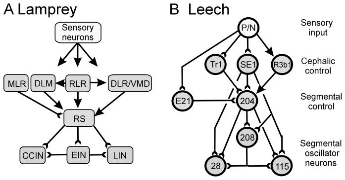
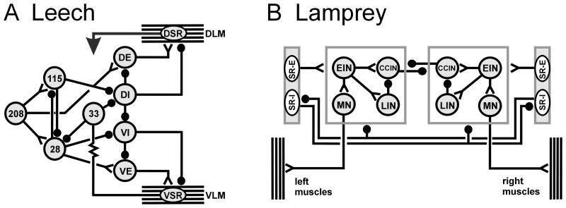
) indicate monosynaptic connections; arrows indicate excitatory polysynaptic pathways that are not identified. RLR – rostrolateral rhombencephalon ; MLR – mesencepalic locomotor region; DLM – dorsolateral mesencepalon ; VMD – ventromedial diencepalon; DLR – diencepalic locomotor region ; RS – reticulospinal.
Figure 8
Figure 8
Excitatory drive. A Excitation to drive swimming is provided in lampreys by reticulospinal neurons (RS; upper trace), leading to prolonged depolarization with superimposed oscillations in motor neurons (MN; middle trace). Many RS neurons oscillate in phase with motor bursts, which is thought to be a result of feedback from spinal neurons. The locomotor activity was initiated by a dimming of the lights. B Injection of a brief (0.22 s) pulse of depolarizing current (third trace) into swim-gating cell 204 (upper trace) can elicit swimming activity that is driven by prolonged cell 204 depolarization and maintains the depolarization of oscillator interneuron IN 28 (second trace). Preparation was superfused with saline containing 50 μM serotonin. IN 28 was slightly hyperpolarized by continuous current injection. VR – ventral nerve root recording; DP – dorsal-posterior nerve recording; (R/L, X), R/L refers to the left or right side, X is the ganglion number.
Figure 9
Figure 9
Intracellular potentials during fictive swimming. A Leech. Membrane potentials in interneurons (IN) 115 and 208 (upper two traces), both swim oscillator interneurons, compared to extracellular motor bursts (bottom trace). Swimming was evoked by brief stimulation (at large artifacts) of a segmental nerve. Because the midpoint of the dorsal-posterior (DP) nerve impulse bursts occur concurrently with the peak of the IN oscillations, they are designated with the same activity phase (0%). B Lamprey. Membrane potentials in a lateral IN (LIN) and a motor neuron (MN) occur phase-locked to MN impulse bursts recorded from a ventral root (VR) in a brainstem-spinal cord preparation. Both LIN and the MN are depolarized during ventral root bursts and hence have a phase of 0%. Swimming activity in the lamprey preparation was elicited by electric shock of the spinal cord (large artifacts).
Figure 10
Figure 10
Neuronal circuits for generating swim oscillations. A Leech circuits. A1 The current minimal model for swim generation is a circuit of three inhibitory INs that form an inhibitory ring. Such a circuit generates oscillations that have three phases without strong dependence on cellular properties. A2 Summary of many of the segmental interactions between MNs and INs. The numbers denote individually identified INs; DI-102 and DI-1 are inhibitory MNs. Note that inhibitory MNs are strongly interconnected with the INs and may contribute significantly to rhythm generation. Phase values for the three columns of neurons in the CPG are indicated at the top. B Lamprey circuits. B1 “Half-center” model for spinal interactions leading to vertebrate locomotion. Two neurons oscillate in anti-phase because of reciprocal inhibitory interactions and because of critical cellular properties. B2 Circuit summary for the segmental CPG in lamprey. Crossed inhibitory interactions ensure that when one side is active, the other is inhibited. Abbreviations: MN, motor neuron; DI, dorsal longitudinal inhibitor; CCIN, contralaterally and caudally projecting interneuron; EIN, excitatory interneuron; LIN, lateral interneuron. Lines ending in filled circles (formula image
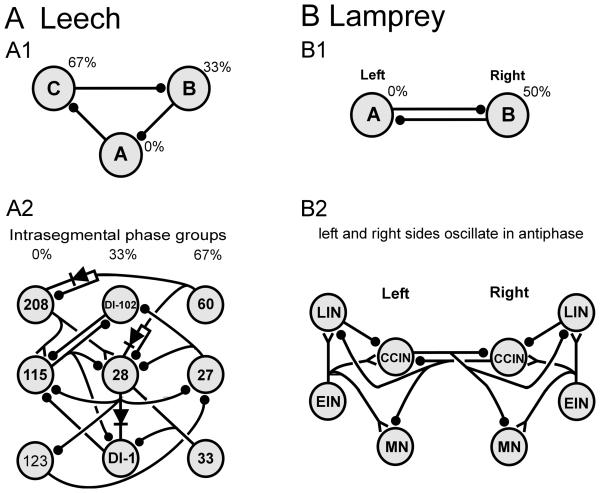
) denote inhibitory synapses; those terminating with a Y (formula image) are excitatory; diode symbols denote rectifying electrical junctions.
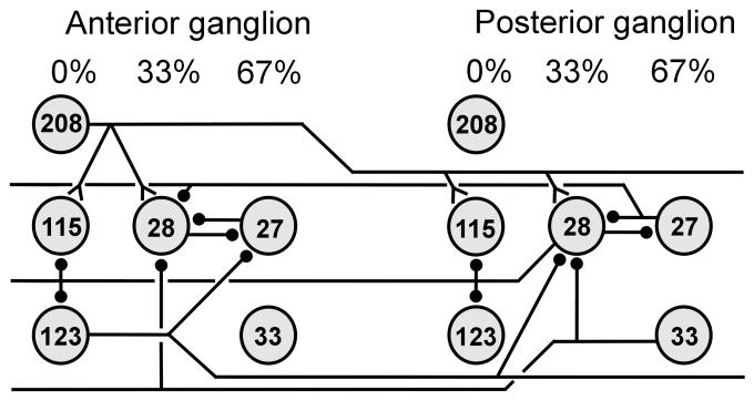
Figure 11
Figure 11
Intersegmental coordinating interactions in the leech. The intersegmental interactions shown extend both in the rostral and caudal directions for about 5 segments. There is only one identified excitatory oscillator neuron, IN 208.. The interactions shown quantitatively account for intersegmental phase lags during fictive swimming. Symbols are as in Fig. 10.
Figure 12
Figure 12
Elimination of long-range interactions. A Lamprey preparation. Long-distance axons in the spinal cord are interrupted through contralateral hemisections separated by several segments. B Leech Z-cut preparation. To interrupt through-going interactions, the right lateral intersegmental connective nerve is cut rostral to ganglion M10 and the left connective is cut caudal to M10. Schematic in A from Guan et al., 2001.
Figure 13
Figure 13
Manipulation of intersegmental phase lags through changes in local, segmental cycle period. A Period changes controlled by saline temperature superfusing the leech nerve cord. Splitting the recording chamber (vertical dashed line) allowed independent control of rostral (TA) and caudal (TP) nerve cord temperature, and hence intrinsic local cycle periods. Intersegmental phase lags were decreased when TA was less than TP and increased when TA was greater than TP. B Period changes controlled by NMDA concentrations in the lamprey spinal cord. The recording chamber was split into three compartments allowing independent control of cycle period in rostral, middle and caudal portions of the spinal cord. Inset shows the recording arrangement for the ventral root traces. The numbers above each set of traces indicate the NMDA concentrations in μM. Decreasing cycle period in caudal segments by elevating NMDA led to a reversal of the normal phase lag, with caudal segments now leading rostral ones. Decreasing cycle period in the middle chamber caused this portion to phase-lead the rostral and caudal compartments. Trace in B is from Matsushima and Grillner (1992).
Figure 14
Figure 14
Form and function of stretch receptors. A Leech. The terminals of stretch receptors in the leech terminate broadly within segmental ganglia. Recordings are from the giant axons near the ganglion edge (VSR electrode). The records in A arose from an experiment in which an excitatory MN (upper trace, VE-4) was excited via intracellular current injection to induce increased tension in a fixed-length flap piece of body wall (bottom trace). The increased isometric tension induced a hyperpolarization in the giant axon of the ventral stretch receptor (VSR, middle trace). B Lamprey. The edge cell (EC) has processes that terminate near the lateral edge of the spinal cord and an ipsilaterally projecting axon. Stretching the margin of the spinal cord depolarizes the edge cell and gives rise to sustained impulse activity. The small upward and downward deflections in the lower trace indicated step increases (stretch) and decreases (release) in length, respectively. A is constructed from Fig. 2 of Cang et al. (2001). B is constructed from Fig. 3 in Grillner et al. (1984).
Figure 15
Figure 15
Interactions of stretch receptors with the CPG. A Leech circuit. Muscle tension is detected by the dorsal (DSR) and ventral (VSR) stretch receptors. The VSR neuron has strong non-rectifying electrical interactions with IN 33 and hence is directly interconnected the CPG. Interactions between the DSR and the segmental neurons are unknown. B Lamprey circuit. There are two classes of stretch receptors (a.k.a., edge cells). One (SR-E) excites most neurons in the ipsilateral CPG. The second (SR-I) makes inhibitory contacts with contralateral CCINs and LINs and inhibits the contralateral SR-I as well. Both types of stretch receptors have processes near the lateral margin of the spinal cord and detect changes in spinal cord length caused either by imposed bending or by contraction of segmental muscles. A is redrawn from Fig. 5, Friesen and Kristan, 2007; B is redrawn from Viana Di Prisco et al. 1990. Note that only a subset of CPG interactions are shown. Symbols are as in Fig. 10. The resistor symbol denotes a nonrectifying electrical connection.
Figure 16
Figure 16
Simplified systems overview. A A cell-to-cell pathway has been identified in leeches from sensory inputs to motor output. B The reticulospinal (RS) spinal system that drives swimming and the oscillator interneurons in the lamprey are relatively well characterized, but other aspects are less well understood. It is not clear what neurons serve trigger functions. Many of the synaptic interactions in the excitatory cascades that drive swimming in leeches (A) and lampreys (B) are mediated by glutamatergic receptors. Neuromodulators and sensory feedback (not shown) are also important to the swim systems. P, pressure cell; Tr1, trigger neuron 1; SE1, swim excitor neuron 1; RZ - Retzius cell ; DE, dorsal excitor ; MLR, mesencephalic locomotor region; RS, reticulospinal cell; CCIN, caudal and contralaterally projecting interneuron; LIN, lateral interneuron; EIN, excitatory interneuron; MN, motor neuron. Symbols as in Fig. 7 and Fig. 10.

References

    1. Antri M, Fenelon K, Dubuc R. The contribution of synaptic inputs to sustained depolarizations in reticulospinal neurons. J. Neurosci. 2009;29:1140–1151. - PMC - PubMed
    1. Archambault PS, Deliagina TG, Orlovsky GN. Non-undulatory locomotion in the lamprey. Neuroreport. 2001;12:1803–1807. - PubMed
    1. Blackshaw SE, Thompson SW. Hyperpolarizing responses to stretch in sensory neurones innervating leech body wall muscle. J. Physiol. 1988;396:121–37. - PMC - PubMed
    1. Bowtell G, Williams TL. Anguilliform body dynamics: modelling the interaction between muscle activation and body curvature. Philos. Trans. R. Soc. Lond. B. Biol. Sci. 1991;334:385–390. - PubMed
    1. Boyd MR, McClellan AD. Changes in locomotor activity parameters with variations in cycle time in larval lamprey. J. Exp. Biol. 2002;205:3707–3716. - PubMed

Publication types

MeSH terms