. 2011 Jan 21;405(3):630-41.
doi: 10.1016/j.jmb.2010.10.043. Epub 2010 Nov 19.

Creating designed zinc-finger nucleases with minimal cytotoxicity

Affiliations

Creating designed zinc-finger nucleases with minimal cytotoxicity

Sivaprakash Ramalingam et al. J Mol Biol. .

Abstract

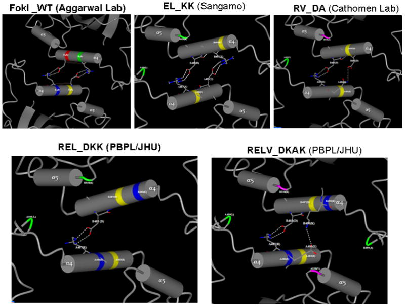
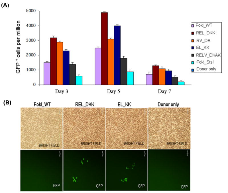
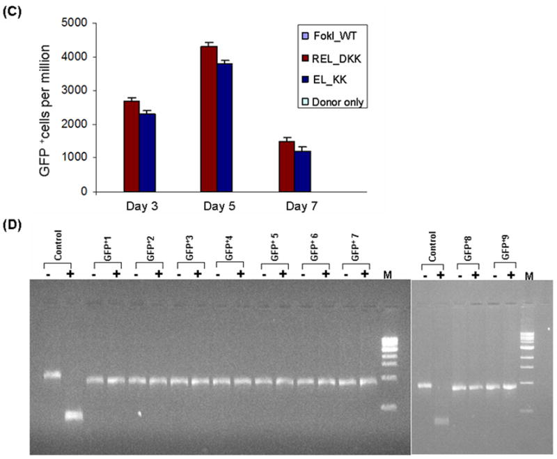
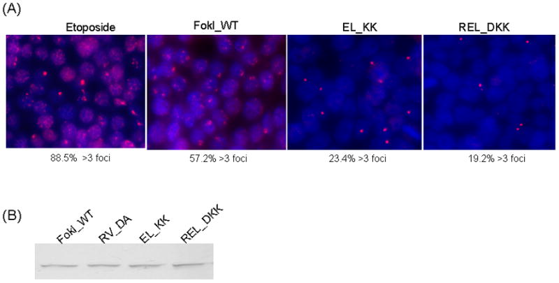
Zinc-finger nucleases (ZFNs) have emerged as powerful tools for delivering a targeted genomic double-strand break (DSB) to either stimulate local homologous recombination with investigator-provided donor DNA or induce gene mutations at the site of cleavage in the absence of a donor by nonhomologous end joining both in plant cells and in mammalian cells, including human cells. ZFNs are formed by fusing zinc-finger proteins to the nonspecific cleavage domain of the FokI restriction enzyme. ZFN-mediated gene targeting yields high gene modification efficiencies (>10%) in a variety of cells and cell types by delivering a recombinogenic DSB to the targeted chromosomal locus, using two designed ZFNs. The mechanism of DSB by ZFNs requires (1) two ZFN monomers to bind to their adjacent cognate sites on DNA and (2) the FokI nuclease domains to dimerize to form the active catalytic center for the induction of the DSB. In the case of ZFNs fused to wild-type FokI cleavage domains, homodimers may also form; this could limit the efficacy and safety of ZFNs by inducing off-target cleavage. In this article, we report further refinements to obligate heterodimer variants of the FokI cleavage domain for the creation of custom ZFNs with minimal cellular toxicity. The efficacy and efficiency of the reengineered obligate heterodimer variants of the FokI cleavage domain were tested using the green fluorescent protein gene targeting reporter system. The three-finger and four-finger zinc-finger protein fusions to the REL_DKK pair among the newly generated FokI nuclease domain variants appear to eliminate or greatly reduce the toxicity of designer ZFNs to human cells.


Figures

Figure 1
Figure 1
Predicted profiles of H-bond interactions between α4 helices at the dimer interface of obligate heterodimer variants of FokI nuclease domains, based on protein modeling and energy minimization. The 3D structure of the FokI dimer was obtained from RCSB Protein Data Bank, which was generated by X-ray diffraction method at a resolution of 2.3 A by Aggarwaal’s lab (36). The CCP4 Molecular Graphics software (Version 6.1.2) for Macromolecular X-ray Crystallography was used for 3D structure analysis and SPDBV software for protein modeling. Only hydrogen bond interactions between α4 helices at the dimer interface are shown. All potential dimer interface interactions (H-bond interactions and hydrophobic interactions) are listed in Table S2.
Figure 2
Figure 2
Testing the efficiency and efficacy of CCR5 3- and 4-finger ZFNs (generated by fusing the corresponding ZFPs to various obligate heterodimer variants of FokI nuclease domain) using the GFP gene targeting reporter system. HEK293 cells carrying a mutated eGFP reporter gene were transiently transfected with a donor plasmid carrying a fragment of wild-type GFP and plasmids expressing various 3- or 4-finger CCR5-specific ZFN constructs, using Lipofectamine 2000 as described elsewhere (21,27). Transfections of various obligate heterodimer variants and FokI_WT were performed one after another on the same day. After each transfection, the treated cells were split into 2 flasks. GFP positive cells in about 10,000 treated cells in each flask were determined by FACS and then normalized to one million treated cells. The difference between the two independent FACS readings is shown as error bars. A) Frequency of gene correction in HEK293 Flp-In cells of a chromosomal mutant GFP reporter disabled by insertion of the CCR5 ZFN target sequences using 4-finger ZFNs (28, 29). Quantitative FACS analyses of the GFP positive cells at 3, 5 and 7 days post-transfection with designer CCR5-specific 4-finger ZFNs (constructs carrying the FokI_WT and obligate heterodimer FokI nuclease domain variants) and donor plasmids. WT: wild type; EL_KK, 4-finger CCR5-ZFNs containing the FokI nuclease domain mutants reported by Miller et al., 2007 (32). RV_DA, 4-finger CCR5-ZFN constructs carrying the FokI nuclease domain mutants reported by Szczepek et al., 2007 (33). REL_DKK, RELV_DKAK and FokI_StsI, 4-finger CCR5-ZFN constructs carrying the FokI nuclease domain mutants that were generated at PBPL/JHU. B) Top panel: HEK293 Flp-In cells 5 days post-transfection as seen in brightfield; Bottom panel: GFP positive cells as seen 5 days post-transfection of HEK293 Flp-In cells with 3-finger CCR5-ZFN constructs (carrying Fok_WT, EL_KK or REL_DKK respectively) and donor plasmid. No GFP positive cells were seen with either donor alone or ZFNs containing FokI_WT nuclease domains and donor plasmid. C) FACS analyses of the frequency of gene correction in HEK293 Flp-In cells using 3-finger CCR5 ZFN constructs carrying FokI_WT, EL_KK, REL_DKK or donor alone, respectively. Results from two independent transfections, performed on different days, are shown in Figure S1; both transfections showed a similar trend of gene correction efficiencies for EL_KK and REL_DKK, respectively. The dose response curve using titrations of 3-finger ZFN expression plasmids, EL_KK and REL_DKK respectively, at constant donor plasmid, are shown in Figure S2. D) Analysis of the genotype of nine different individual GFP positive clones. Five days post-transfection with CCR5 3-finger ZFNs and the donor plasmids, GFP positive cells were sorted, serially diluted to get individual clones and grown. The genomic DNA was isolated from the GFP positive clones and the eGFP gene at the Flp-In locus was PCR amplified and digested with BstXI. The mutant eGFP gene has two BstXI sites, where the ZFN binding sites are inserted. Correction of the eGFP gene by homology-directed repair results in the loss of the BstXI sites. The PCR product size of the corrected eGFP gene is 930 bp as compared to 990 bp for the mutant gene. BstXI digestion of the mutant eGFP PCR product generates two bands: 450 bp and 540 bp, respectively. Lanes: Control, PCR product of the mutant eGFP gene from untransfected cells before (-) and after (+) digestion with BstXI; GFP+1–9, PCR products of 9 different individual clones obtained from GFP positive sorted cells before (-) and after (+) digestion with BstXI; M, 1 Kb ladder. All GFP positive cells are resistant to BstXI digestion, confirming ZFN-mediated eGFP gene correction in these cells.
Figure 2
Figure 2
Testing the efficiency and efficacy of CCR5 3- and 4-finger ZFNs (generated by fusing the corresponding ZFPs to various obligate heterodimer variants of FokI nuclease domain) using the GFP gene targeting reporter system. HEK293 cells carrying a mutated eGFP reporter gene were transiently transfected with a donor plasmid carrying a fragment of wild-type GFP and plasmids expressing various 3- or 4-finger CCR5-specific ZFN constructs, using Lipofectamine 2000 as described elsewhere (21,27). Transfections of various obligate heterodimer variants and FokI_WT were performed one after another on the same day. After each transfection, the treated cells were split into 2 flasks. GFP positive cells in about 10,000 treated cells in each flask were determined by FACS and then normalized to one million treated cells. The difference between the two independent FACS readings is shown as error bars. A) Frequency of gene correction in HEK293 Flp-In cells of a chromosomal mutant GFP reporter disabled by insertion of the CCR5 ZFN target sequences using 4-finger ZFNs (28, 29). Quantitative FACS analyses of the GFP positive cells at 3, 5 and 7 days post-transfection with designer CCR5-specific 4-finger ZFNs (constructs carrying the FokI_WT and obligate heterodimer FokI nuclease domain variants) and donor plasmids. WT: wild type; EL_KK, 4-finger CCR5-ZFNs containing the FokI nuclease domain mutants reported by Miller et al., 2007 (32). RV_DA, 4-finger CCR5-ZFN constructs carrying the FokI nuclease domain mutants reported by Szczepek et al., 2007 (33). REL_DKK, RELV_DKAK and FokI_StsI, 4-finger CCR5-ZFN constructs carrying the FokI nuclease domain mutants that were generated at PBPL/JHU. B) Top panel: HEK293 Flp-In cells 5 days post-transfection as seen in brightfield; Bottom panel: GFP positive cells as seen 5 days post-transfection of HEK293 Flp-In cells with 3-finger CCR5-ZFN constructs (carrying Fok_WT, EL_KK or REL_DKK respectively) and donor plasmid. No GFP positive cells were seen with either donor alone or ZFNs containing FokI_WT nuclease domains and donor plasmid. C) FACS analyses of the frequency of gene correction in HEK293 Flp-In cells using 3-finger CCR5 ZFN constructs carrying FokI_WT, EL_KK, REL_DKK or donor alone, respectively. Results from two independent transfections, performed on different days, are shown in Figure S1; both transfections showed a similar trend of gene correction efficiencies for EL_KK and REL_DKK, respectively. The dose response curve using titrations of 3-finger ZFN expression plasmids, EL_KK and REL_DKK respectively, at constant donor plasmid, are shown in Figure S2. D) Analysis of the genotype of nine different individual GFP positive clones. Five days post-transfection with CCR5 3-finger ZFNs and the donor plasmids, GFP positive cells were sorted, serially diluted to get individual clones and grown. The genomic DNA was isolated from the GFP positive clones and the eGFP gene at the Flp-In locus was PCR amplified and digested with BstXI. The mutant eGFP gene has two BstXI sites, where the ZFN binding sites are inserted. Correction of the eGFP gene by homology-directed repair results in the loss of the BstXI sites. The PCR product size of the corrected eGFP gene is 930 bp as compared to 990 bp for the mutant gene. BstXI digestion of the mutant eGFP PCR product generates two bands: 450 bp and 540 bp, respectively. Lanes: Control, PCR product of the mutant eGFP gene from untransfected cells before (-) and after (+) digestion with BstXI; GFP+1–9, PCR products of 9 different individual clones obtained from GFP positive sorted cells before (-) and after (+) digestion with BstXI; M, 1 Kb ladder. All GFP positive cells are resistant to BstXI digestion, confirming ZFN-mediated eGFP gene correction in these cells.
Figure 3
Figure 3
Reduced genome-wide DNA damage levels by REL_DKK obligate heterodimer variant pair of FokI nuclease domain. A), Representative images of cells treated with the DNA cleavage agent etoposide or transfected with the indicated ZFN expression plasmids. Cells were fixed after 30 h and stained with antibodies against 53BP1 (red) and then with DAPI (blue). The fraction of cells containing more than 3 foci is indicated under each panel. The total number of cells analyzed is: etoposide, 152; FokI_WT, 245; EL_KK, 282; REL_DKK, 289. B), ZFN expression levels were examined by anti-FokI immunoblot analysis. HEK293 Flp-In cells were transfected with indicated ZFN expression plasmids and cells were harvested after 30 h. Equal amounts of total cellular protein was separated by 10% SDS-PAGE gel and transferred to PVDF membrane. The blot was probed with anti-FokI antibody. The ZFNs migrate as a single band on the gel. Western blot analysis shows comparable levels ZFN expression for various obligate heterodimer variants in HEK293 Flp-In cells.

Similar articles

Cited by

References

    1. Kim YG, Cha J, Chandrasegaran S. Proc Natl Acad Sci USA. 1996;93:1156–1160. - PMC - PubMed
    1. Li L, Wu LP, Chandrasegaran S. Proc Natl Acad Sci USA. 1992;89:4275–4279. - PMC - PubMed
    1. Lin L, Chandrasegaran S. Proc Natl Acad Sci USA. 1993;90:2764–2768. - PMC - PubMed
    1. Kim YG, Chandrasegaran S. Proc Natl Acad Sci USA. 1994;91:883–887. - PMC - PubMed
    1. Bibikova M, Carroll D, Segal DJ, Trautman JK, Smith J, Kim Y–G, Chandrasegaran S. Mol Cell Biol. 2001;21:289–297. - PMC - PubMed

Publication types

MeSH terms

Substances

LinkOut - more resources