

Impairment of IGF-I expression and anabolic signaling following ischemia/reperfusion in skeletal muscle of old mice
- PMID: 21094246
- PMCID: PMC3061979
- DOI: 10.1016/j.exger.2010.11.002
Impairment of IGF-I expression and anabolic signaling following ischemia/reperfusion in skeletal muscle of old mice
Abstract
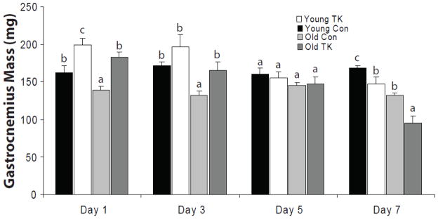
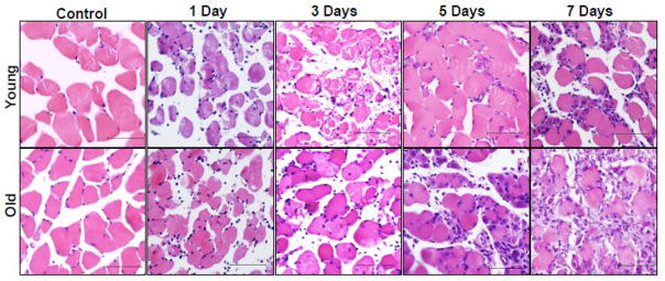
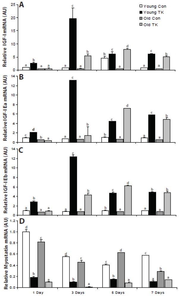
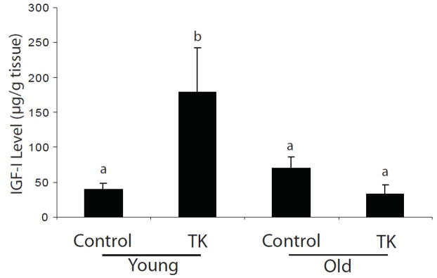
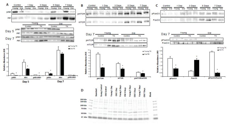
With the advancement of age, skeletal muscle undergoes a progressive decline in mass, function, and regenerative capacity. Previously, our laboratory has reported an age-reduction in recovery and local induction of IGF-I gene expression with age following tourniquet (TK)-induced skeletal muscle ischemia/reperfusion (I/R). In this study, young (6 mo) and old (24-28 mo) mice were subjected to 2h of TK-induced ischemia of the hindlimb followed by 1, 3, 5, or 7 days of reperfusion. Real time-PCR analysis revealed clear age-related reductions and temporal alterations in the expression of IGF-I and individual IGF-I Ea and Eb splice variants. ELISA verified a reduction of IGF-I peptide with age following 7 day recovery from TK. Western blotting showed that the phosphorylation of Akt, mTOR, and FoxO3, all indicators of anabolic activity, were reduced in the muscles of old mice. These data indicate that an age-related impairment of IGF-I expression and intracellular signaling does exist following injury, and potentially has a role in the impaired recovery of skeletal muscle with age.
Copyright © 2010 Elsevier Inc. All rights reserved.
Figures
Similar articles
-
Functional deficits and insulin-like growth factor-I gene expression following tourniquet-induced injury of skeletal muscle in young and old rats.J Appl Physiol (1985). 2008 Oct;105(4):1274-81. doi: 10.1152/japplphysiol.90418.2008. Epub 2008 Jul 31. J Appl Physiol (1985). 2008. PMID: 18669936 Free PMC article.
-
Testosterone regulation of Akt/mTORC1/FoxO3a signaling in skeletal muscle.Mol Cell Endocrinol. 2013 Jan 30;365(2):174-86. doi: 10.1016/j.mce.2012.10.019. Epub 2012 Oct 29. Mol Cell Endocrinol. 2013. PMID: 23116773 Free PMC article.
-
Protective Effects of Sonic Hedgehog Against Ischemia/Reperfusion Injury in Mouse Skeletal Muscle via AKT/mTOR/p70S6K Signaling.Cell Physiol Biochem. 2017;43(5):1813-1828. doi: 10.1159/000484068. Epub 2017 Oct 19. Cell Physiol Biochem. 2017. PMID: 29065414
-
Longevity and skeletal muscle mass: the role of IGF signalling, the sirtuins, dietary restriction and protein intake.Aging Cell. 2015 Aug;14(4):511-23. doi: 10.1111/acel.12342. Epub 2015 Apr 10. Aging Cell. 2015. PMID: 25866088 Free PMC article. Review.
-
Resistance training, and IGF involvement in the maintenance of muscle mass during the aging process.Ageing Res Rev. 2006 Aug;5(3):310-31. doi: 10.1016/j.arr.2006.05.001. Epub 2006 Sep 1. Ageing Res Rev. 2006. PMID: 16949353 Review.
Cited by
-
Controlled release of IGF-I from a biodegradable matrix improves functional recovery of skeletal muscle from ischemia/reperfusion.Biotechnol Bioeng. 2012 Apr;109(4):1051-9. doi: 10.1002/bit.24382. Epub 2011 Dec 1. Biotechnol Bioeng. 2012. PMID: 22095096 Free PMC article.
-
Mice producing reduced levels of insulin-like growth factor type 1 display an increase in maximum, but not mean, life span.J Gerontol A Biol Sci Med Sci. 2014 Apr;69(4):410-9. doi: 10.1093/gerona/glt108. Epub 2013 Jul 20. J Gerontol A Biol Sci Med Sci. 2014. PMID: 23873963 Free PMC article.
-
Controlled delivery of SDF-1α and IGF-1: CXCR4(+) cell recruitment and functional skeletal muscle recovery.Biomater Sci. 2015 Nov;3(11):1475-86. doi: 10.1039/c5bm00233h. Biomater Sci. 2015. PMID: 26247892 Free PMC article.
-
Interaction between insulin-like growth factor-1 and atherosclerosis and vascular aging.Front Horm Res. 2014;43:107-24. doi: 10.1159/000360571. Epub 2014 Jun 10. Front Horm Res. 2014. PMID: 24943302 Free PMC article. Review.
-
Expression of insulin-like growth factor 1 isoforms in the rabbit oculomotor system.Growth Horm IGF Res. 2011 Aug;21(4):228-32. doi: 10.1016/j.ghir.2011.06.001. Epub 2011 Jun 23. Growth Horm IGF Res. 2011. PMID: 21703892 Free PMC article.
References
-
- Adamo ML, Farrar RP. Resistance training, and IGF involvement in the maintenance of muscle mass during the aging process. Ageing Res Rev. 2006;5:310–31. - PubMed
-
- Conboy IM, Rando TA. Aging, stem cells and tissue regeneration: lessons from muscle. Cell Cycle. 2005;4:407–10. - PubMed
-
- Zerba E, Komorowski TE, Faulkner JA. Free radical injury to skeletal muscles of young, adult, and old mice. Am J Physiol. 1990;258:C429–35. - PubMed
-
- Brooks SV, Faulkner JA. Contraction-induced injury: recovery of skeletal muscles in young and old mice. Am J Physiol. 1990;258:C436–42. - PubMed
Publication types
MeSH terms
Substances
Grants and funding
LinkOut - more resources
Full Text Sources
Medical
Research Materials
Miscellaneous
