

Experimental infection of dogs (Canis familiaris) with sporulated oocysts of Neospora caninum
- PMID: 21094584
- PMCID: PMC7116961
- DOI: 10.1016/j.vetpar.2010.10.047
Experimental infection of dogs (Canis familiaris) with sporulated oocysts of Neospora caninum
Abstract
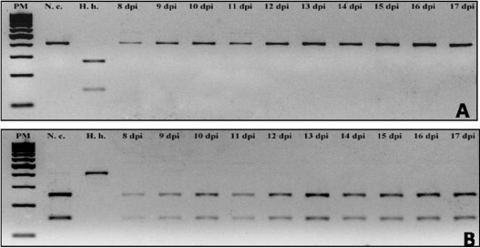
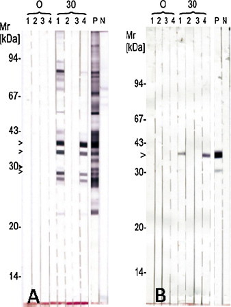
Neospora caninum is widely distributed in the world and this parasite is one of the major causes of abortion in cattle. Dogs and coyotes are definitive hosts of N. caninum and several species of domestic and wild animals are intermediate hosts. Dogs can become infected by the ingestion of tissues containing cysts and then excrete oocysts. It is not yet known whether sporulated oocysts are able to induce a patent infection in dogs, i.e. a shedding of N. caninum oocysts in feces. The objective of this study was to experimentally examine the infection of dogs by sporulated oocysts. The oocysts used in the experiment were obtained by feeding dogs with brain of buffaloes (Bubalus bubalis) positive for anti-N. caninum antibodies by indirect fluorescent antibody test (IFAT ≥200). Oocysts shed by these dogs were confirmed to be N. caninum by molecular methods and by bioassay in gerbils, and sporulated N. caninum oocysts were used for the oral infection of four dogs. The dogs were 8 weeks old and negative for antibodies to N. caninum and Toxoplasma gondii. Dogs 1 and 4 received an inoculum of 10,000 sporulated oocysts each; dog 2 an inoculum of 5000 sporulated oocysts and dog 3 received 1000 sporulated oocysts of N. caninum. The total feces excreted by these dogs were collected and examined daily for a period of 30 days. No oocysts were found in their feces. The dogs were monitored monthly for a 6-month period to observe a possible seroconversion and when this occurred the animals were eliminated from the experiment. Dogs 1 and 4 seroconverted 1 month after the infection with titer, in the IFAT, of 1600 and 800, respectively; the other two dogs presented no seroconvertion during the 6-month period. Dogs 1 and 2 were euthanized 180 days after infection and were examined for the detection of N. caninum in tissues (brain, muscle, lymph node, liver, lung, heart and bone marrow) by immunohistochemistry and PCR with negative results in both techniques. Bioassay in gerbils with brain of these dogs was also performed and again the results were negative. In conclusion, dogs infected with sporulated oocysts of N. caninum were not able to shed oocysts in feces. However, a higher dose of infection stimulated the production of antibodies against N. caninum in the dogs.
Copyright © 2010 Elsevier B.V. All rights reserved.
Figures
References
-
- Anderson M.L., Blanchard P.C., Barr B.C., Dubey J.P., Hoffman R.L., Conrad P.A. Neospora-like protozoan infection as a major cause of abortion in California dairy cattle. Journal of American Veterinary Medical Association. 1991;198:241–244. - PubMed
-
- Basso W., Venturini L., Venturini M.C., Hill D.E., Kwok O.C.H., Shen S.K., Dubey J.P. First isolation of Neospora caninum from the feces of a naturally infected dog. Journal of Parasitology. 2001;87:612–618. - PubMed
-
- Basso W., Herrmann D.C., Conraths F.J., Pantchev N., Vrhovec M.G. First isolation of Neospora caninum from the faeces of a dog from Portugal. Veterinary Parasitology. 2009;159:162–166. - PubMed
-
- Bjerkås I., Mohn S.F., Presthus J. Unidentified cyst-forming sporozoon causing encephalomyelitis and myositis in dogs. Zeitschrift für Parasitenkunde. 1984;70:271–274. - PubMed
-
- Cheadle M.A., Lindsay D.S., Blagburn B.L. Prevalence of antibodies to Neospora caninum in dogs. Veterinary Parasitology. 1999;85:325–330. - PubMed
Publication types
MeSH terms
LinkOut - more resources
Full Text Sources
